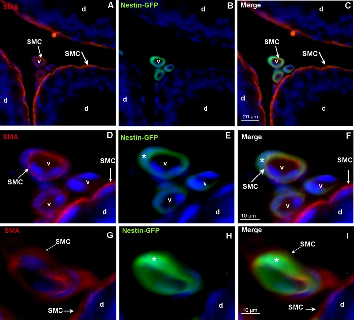
Fig 3. Colocalization of nestin-GFP and the SMC marker SMA in blood vessels.
A-F, G-I: Different vessels (v) of the nestin-GFP mouse. The epididymis (caput) shows nestin-GFP-positive/SMA-positive SMCs. SMCs are indicated. DAPI (blue) labels the nuclei. A,C,D,F,G,I: SMA-positive SMCs of the vessels (v) and of the epididymal duct (d). D-F: Higher magnification of the vessels shown in A-C. C,F,I: Nestin-GFP-positive (asterisks) SMCs in vessels (v).

