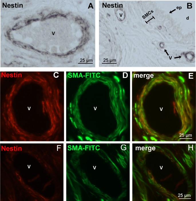
Fig 7. Nestin expression in vessels of the human epididymis.
A,B: Paraffin sections (cauda) with nestin-positive cells in the blood vessels (v), but not in SMCs and epithelial cells (ep) of the epididymal duct (d). C-E, F-H: Immunofluorescence staining of two different vessels with antibodies against nestin (red) and SMA FITC (green). E,H: Merged image. It indicates the localization of nestin in vascular SMCs.

