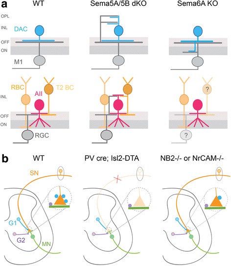
Fig. 2.

Molecular cues guide partner selection of inhibitory neurons. a Schematic showing the lamination of GABAergic-dopaminergic amacrine cells (DACs) and glycinergic AII amacrine cells together with their synaptic partners in wildtype (WT), Sema5A/6A double knockout mutants (dKO) and Sema6A knockouts (KO). T2 BC: Type 2 bipolar cell, M1: melanopsin-expressing retinal ganglion cell, RBC: rod bipolar cell, RGC: retinal ganglion cell, ON: inner sublamina of the retinal plexiform layer, OFF: outer sublamina of the retinal plexiform layer, INL: inner nuclear layer, OPL: outer plexiform layer. Summarized from [18, 19]. Question mark indicates non-examined synaptic partners. b Organization of inhibitory connections in the spinal cord sensory-motor circuit. Distinct populations of inhibitory neurons (G1 and G2) target sensory afferent terminals (SN) and motor neurons (MN), respectively, in WT mice. When sensory afferents are eliminated in PV cre/Isl2-DTA mice, G1 neurons do not form aberrant connections with motor neurons. Inhibitory synapses from G2 to motor neurons are still present in these mutants. In NB2−/− or NrCAM−/− mice, the number of inhibitory synapses from G1 to sensory neurons is significantly reduced but G2 interneuronal contacts onto motor neurons remain unaffected. G1: GABAergic neurons; G2: GABAergic and/or glycinergic neurons. Summarized from [21, 22]
