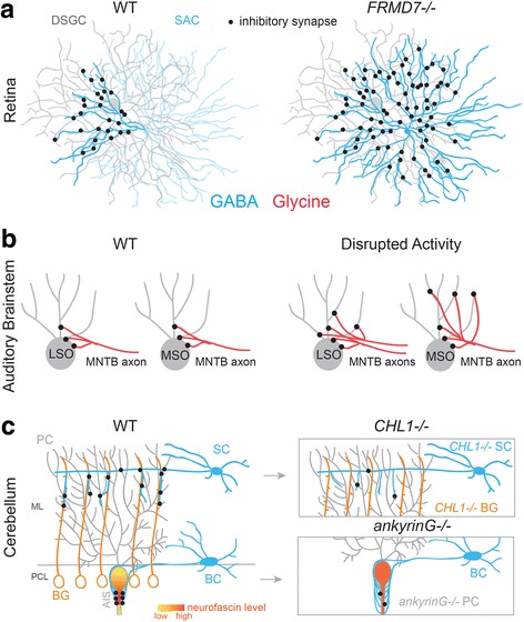
Fig. 3.

Mechanisms that regulate pre- and postsynaptic subcellular targeting of inhibitory connections. a In wildtype (WT) mouse retina, only a specific quadrant of the arbor of GABAergic starburst amacrine cells (SACs) form inhibitory synapses onto direction-selective retinal ganglion cells (DSGCs). In FRMD7−/− mice, this pattern of connectivity between SACs and DSGCs that prefer horizontal movement is disrupted. Summarized from [25]. b During normal development, excess MNTB axon targeting individual LSO neurons are eliminated. In the gerbil auditory brainstem, MNTB neurons initially provide inhibition to MSO neurons across their soma and dendritic arbor, but during development, dendritic synapses are eliminated after the onset of binaural input. Disrupted activity, such as loss of glutamate release or disrupted binaural input, prevents synapse elimination during development. Summarized from: [28, 117, 134–137]. c In the cerebellum, GABAergic stellate cells (SC) and basket cells (BC) utilize distinct cellular mechanisms to target distal dendrites and axon initial segments (AIS) of Purkinje cells (PC). In WT mice, ankyrinG binds to neurofascin and both are highly expressed in the AISs of PCs. Accordingly, in ankyrinG−/− mice the expression pattern of neurofascin is disrupted and basket cell processes erroneously target PC soma and distal processes, following the perturbed neurofascin expression pattern. The number of inhibitory synapses from basket cell to PC AISs is also reduced. In wildtype mice, stellate cells follow processes of Bergmann glia (BG) to make contact with distal dendrites of PCs. Both SCs and BGs express the cell surface molecule (CHL1). Consequently, in CHL1−/− mice stellate cells cannot recognize processes of BG and the number of SC synapses onto PC distal dendrites is reduced. Summarized from [33, 36]. ML: Molecular layer; PCL: Purkinje cell layer
