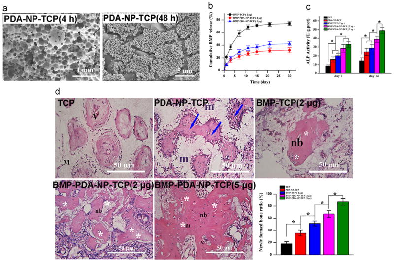
Fig. 6.
PDA nanoparticles (PDA-NPs) for functionalization of porous scaffolds enhance tissue regeneration. a) SEM images of PDA-NPs immobilized onto β-tricalcium phosphate (TCP) scaffolds after incubation in the PDA-NP suspension for 4 h and 48 h. b) Release profiles of bone morphogenetic protein 2 (BMP-2) from various scaffolds in phosphate-buffered saline (PBS) solution. c) ALP activities of BMSCs cultured on various scaffolds for 7 and 14 days. d) Hematoxylin and eosin (H&E) staining images of various scaffolds retrieved after 12-week implantation and quantitative evaluation of newly formed bone on various scaffolds. m: material; nb: new bone; v- vessel; white asterisk: osteocyte; blue arrow: woven bone formation. Statistically significant differences are marked as follows: * for p ≤ 0.05. Adapted and reproduced from Ref. [80].

