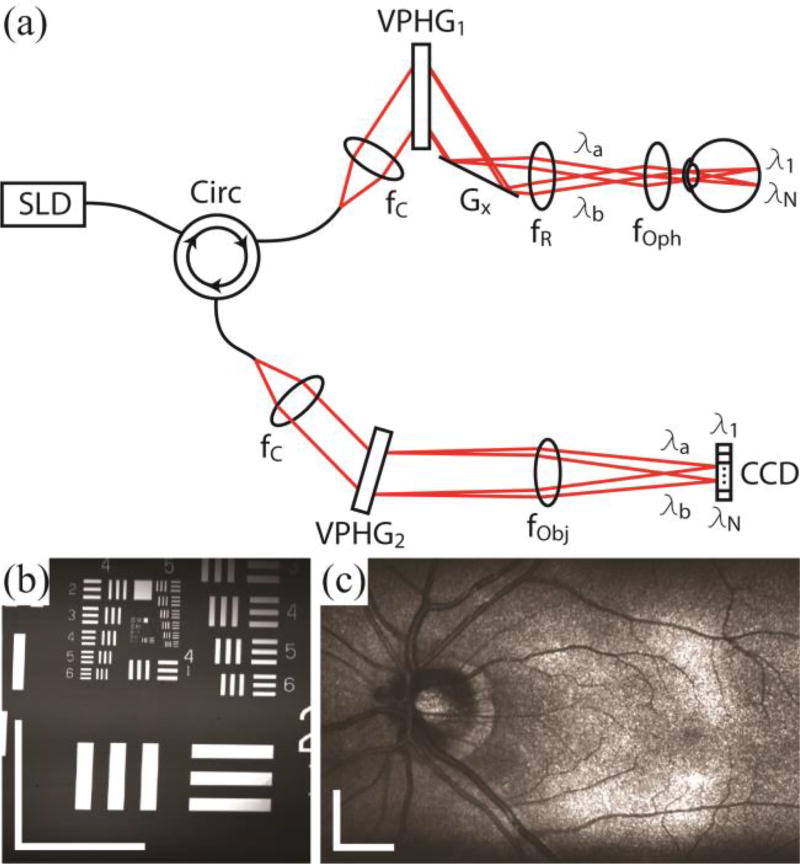
Fig. 1.
Optical schematic and image quality of the SECSLO. (a) Fiber-based spectrally encoded ophthalmoscope. CCD, linear CCD array; f, focal length of collimating, relay, and focusing elements; G, galvanometer; VPHG, grating. (b) 2.5 × 2.5 mm image of USAF 1951 test chart. Spatial resolution was determined to be 16 µm in both lateral directions. (c) 7 × 5 mm (lateral × spectral) image of an average of 5 registered frames of in vivo human fundus. All images are acquired with 1024 × 1024 pixels at 52 kHz line-rate. Illumination power = 700 µW, scale bar = 5 deg.

