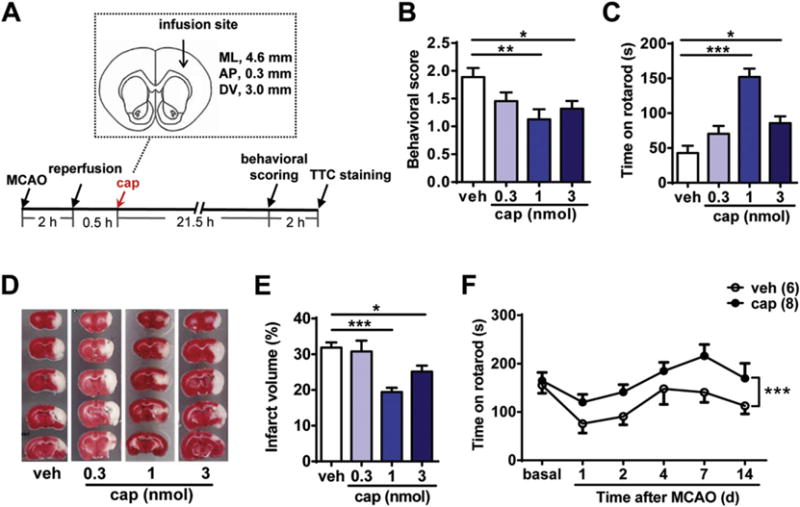
Fig. 1.

Topical application of capsaicin in the peri-infarct area alleviates the ischemic brain injury in the rat MCAO/reperfusion model. (A) Schematic diagram of rat MCAO/reperfusion procedure and capsaicin (cap) infusion site (relative to Bregma). Capsaicin was infused into the S1 area of the cortex. ML, mediolateral; AP, anteroposterior; DV, dorsoventral. (B) Intracortical injection of capsaicin (1 or 3 nmol) improved the neurological scoring of MCAO rats 22 h after reperfusion. n = 8–11, *p < 0.05, **p < 0.01, one-way ANOVA with Dunn’s post hoc tests. veh, vehicle. (C) Intracortical injection of capsaicin (1 or 3 nmol) increased the latency to fall from the accelerated rotarod of MCAO rats 22 h after reperfusion. n = 7–11, *p < 0.05, ***p < 0.001, one-way ANOVA with Dunnett’s post hoc tests. (D) and (E) Intracortical injection of capsaicin (1 or 3 nmol) 0.5 h after reperfusion significantly reduced the cerebral infarct volume. (D) Representative images of TTC-stained brain sections of MCAO/reperfusion rats with injection of different doses of capsaicin (0.3, 1, and 3 nmol) or vehicle. (E) Quantification of the infarct volume in the vehicle- or capsaicin-injected groups. n = 9–11, *p < 0.05, ***p < 0.001, one-way ANOVA with Dunn’s post hoc tests. (F) The improvement of motor coordination function of MCAO rats by capsaicin (1 nmol) persisted for at least 2 weeks. Open circles indicate the vehicle group and solid circles indicate the capsaicin group. ***p < 0.001, two-way ANOVA.
