
Figure 1. HDAC inhibitor treatment does not affect initial recruitment of monocytes and macrophages to the ischemic myocardium.
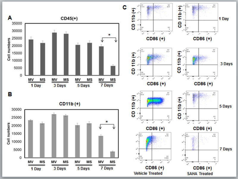
Cell suspensions from infarct zone of vehicle (MV) and SAHA (MS) treated CD1 mice post MI were stained with anti-CD45, CD11b and CD86 mAbs. Results were first processed with live/dead assay and gated with live cells normalized to 200,000 cells. Relative cell numbers are shown as the mean +/− the SEM. A) Live cells were then gated with CD45 positive population to isolate leukocytes. B) Monocytes were then gated with CD11b positive population to identify monocytes [CD45(+)/CD11b(+)]. Any population lower than 103 for either antibody will not be recognized as valid results. C) Original flow cytometry dot plots for infarct tissue in vehicle and SAHA treated mice 1, 3, and 5 and 7 day post-MI. Macrophage were then gated into CD86 positive population that represents classical inflammatory M1 macrophages and will be in region Q2 [CD11b (+)/CD86 (+)] that are greater than 103 for either antibody. (n= 6 for 1, 3, and 5 day groups, n=5 for 7 day group.). *p<0.05 by one-way ANOVA and Bonferroni post-test.