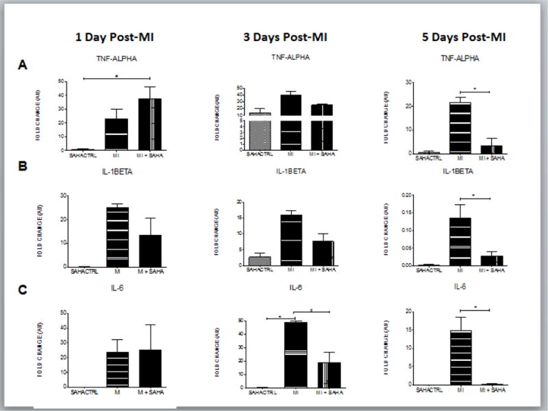
Figure 3. M1 macrophage markers are unchanged at day 1 but drop dramatically at day 5 post-MI with SAHA treatment.

qRT-PCR analysis of mRNA fold change of the M1 associated genes, A) TNF-alpha, B) IL-1beta and C) IL-6 at 1, 3 and 5 days post-MI. Values of all qRT-PCR data are normalized to GAPDH. Each bar represents the fold change +/− SEM of three independent experiments with a group of at least n=3 animals per treatment. #p<0.05 vs control, *p<0.05 vs MI, by one-way ANOVA and Bonferroni post-test.
