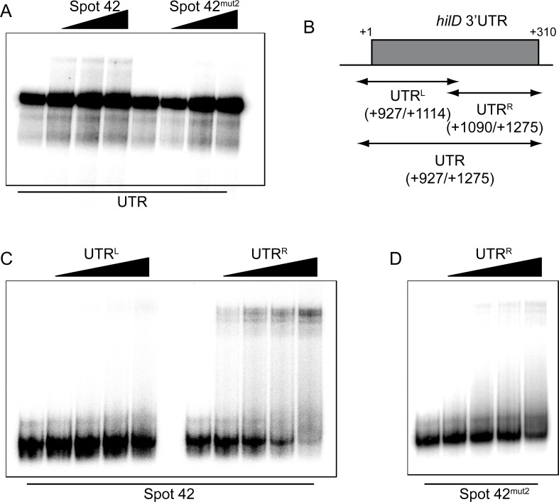
Fig 7. Spot 42 interacts with the downstream part of the hilD 3’UTR.
(A) EMSA assay using 4 nM of hilD 3’UTR RNA radiolabeled incubated with increasing concentration of either Spot 42 or Spot 42mut2 RNA (0, 280, 560, 1700 nM). (B) Schematic representation of the hilD 3’UTR and the UTRL and UTRR fragments. EMSA using 4 nM of radiolabeled Spot 42WT (C) or Spot 42mut2 (D) incubated with increasing concentrations (0, 56, 280, 560, 1,700 nM) of UTRL or UTRR. All RNA transcripts used were obtained by T7 in vitro transcription. Samples were subjected to electrophoresis in a native gel and band shifts were observed upon drying and exposure of the gel.

