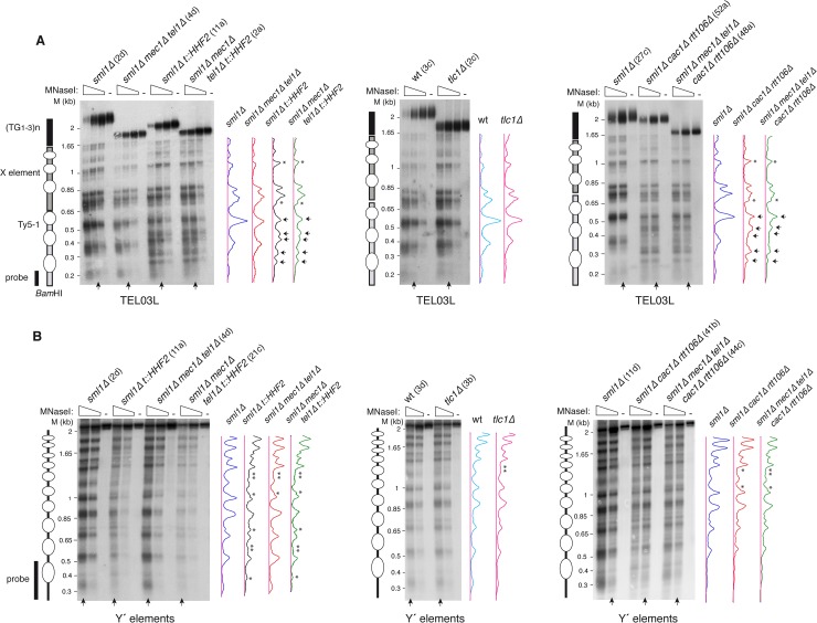
Fig 3. Chromatin analyses of subtelomeric elements in mec1Δ tel1Δ and tlc1Δ pre-senescent cells.
Nucleosome positioning at the left telomere of chromosome III (TEL03L) (A) and global nucleosome pattern at the long subtelomeric Y′ elements (B) from streak 2-derived cultures of the indicated strains. The profiles of MNaseI accessibility of the indicated lanes (vertical arrow), which display similar MNase digestion, are shown on the right. Subtle (asterisks) and major (arrows) changes in chromatin structure are marked in the profiles. Schemes with the position of nucleosomes from the BamHI site at the Ty5-1 element to the end of TEL03L (A), and the bulk nucleosome pattern of the ~2.3 Kb ClaI-ClaI fragment from the long Y′ elements (B) are shown on the left. Note that the analysis in (A) shows nucleosome positioning from a specific site at a single telomere, whereas the analysis in (B) shows the pattern of nucleosomes at and around the probe from all long Y′ elements. Ovals indicate the putative nucleosomes inferred from the MNaseI digestion analysis.

