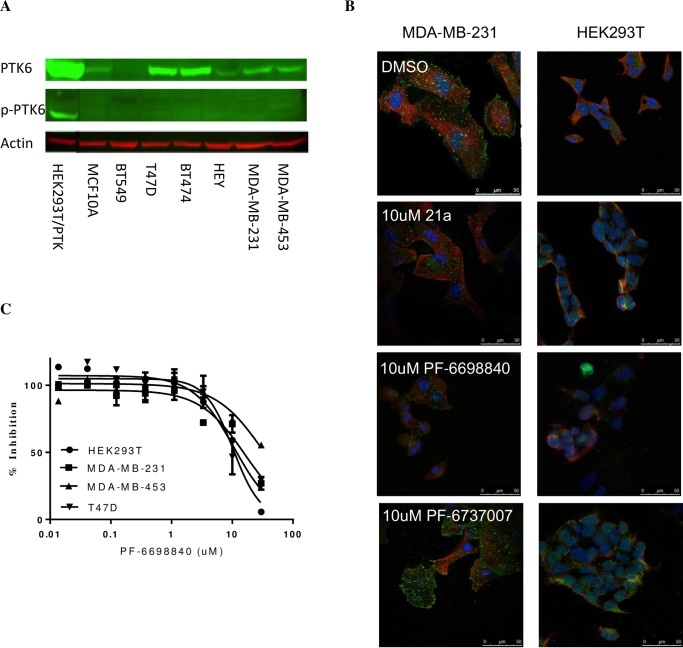
Fig 3. Inhibition of tumor cell growth by Type I and Type II PTK6 inhibitors is independent of PTK6 expression levels in cells.
(A) PTK6 expressions and activation (p-Y342 PTK6) in tumor cell lines and breast epithelial cells MCF10A cells analyzed by Western Blot. Engineered HEK293T cells overexpressing PTK6 WT was used as a positive control. (B) Detection of p-Y342 PTK6 in tumor cells MDA-MB-231 by confocal microscopy. Cells were treated with DMSO, 10 uM PTK6 inhibitor 21a, 10 uM PF-6698840 or 10 uM PTK6-negative control compound PF-6737007 for 2 hours. α-Tubulin (ubiquitously expressed in cells) and DAPI (restricted expression in nucleus) are shown in red and blue, respectively. p-PTK6 (green) was detected on the cell membrane of MDA-MB-231 cells. HEK293T cells do not have detectable PTK6 expression and were used as a negative control. (C) Cell growth inhibition by PTK6 inhibitors in PTK6-positive tumor cells and PTK6-negative HEK293T cells. Cells were treated with DMSO or compounds for 6 days in 2D or 7 days in 3D culture, and the cell growth inhibition was measured by Cell Titer-Glo on day 6 or 7.

