



Abstract
Objectives:
Third molars often require surgical removal. Since three-dimensional radiological assessment is often indicated in difficult cases to avoid surgical complications, the radiation burden has to be considered. Here, MRI may offer a dose-free alternative to conventional X-ray techniques. The aim of this retrospective analysis was to evaluate the assessment quality of MRI compared to panoramic radiography in impacted and partially impacted lower third molars.
Methods:
Panoramic radiographs and MRI scans of 28 Caucasian patients were assessed twice by four investigators. Wisdom teeth were classified according to Juodzbalys and Daugela 2013.
Results:
When radiological lower third molar assessments with panoramic radiography and MRI were compared, staging concurred in 73% in the first round of assessments and 77% in the second.
Conclusions:
The presented study demonstrates that MRI not only provides much the same information that panoramic radiography usually does, but also has the advantages of a dose-free three-dimensional view. This may facilitate and shorten third molar surgery. Image interpretation, however, can differ depending on training and experience.
Introduction
Third molars are frequently extracted, especially in adolescent patients.1 Frequent indications are caries, pericoronitis, or orthodontic problems.2 The difficulty of surgical removal depends on surrounding bone thickness, depth of impaction, angulation, root formation, and close relation to the second molar as well as to the inferior alveolar nerve.3 Treatment planning includes radiological assessment, usually panoramic radiographs, but these two-dimensional dental X-rays yield only limited information.4 Accordingly, cross-sectional imaging, preferably by cone beam CT (CBCT) can be indicated for the prevention of intra- and postoperative complications in difficult cases.5–7 Cross-sectional imaging provides submillimeter accuracy but involves an approximately 10-fold greater dose of ionizing radiation than compared with panoramic radiographs.7, 8 Despite the obvious benefits of these methods, it has to be considered that the patient’s radiation burden is increased with each additional X-ray scan.5, 9 This is especially significant in adolescents, who are far more sensitive to radiation-induced cancer than adults.10
MRI is a well-established dose-free technique with excellent tissue contrast. In spite of its wide use in general medicine, there are as yet only a few applications for dentistry, although recent years have seen increasing interest in MRI as a reliable imaging method in dentistry, suggesting that it could become an alternative to CBCT and CT scans in third molar evaluation.11–14
The aim of this retrospective analysis was to evaluate the assessment quality of MRI compared to panoramic radiography (PR) in impacted and partially impacted lower third molars and to assess whether dental MRI is a potential alternative to CBCT and CT for advanced radiological preoperative assessment.
Methods and materials
The study protocol was approved by the Ethics Committee of the Medical University Graz (EK Nr. 23–353 ex 10/11).
28 Caucasian patients (19 female, 9 male) were included in the study. In a previous study with the same patients,14 panoramic X-rays and MRI scans were used to estimate patients’ ages based on third molar assessment. According to the study protocol of Baumann et al,14 participants had been recruited randomly from patients presenting for surgical removal of impacted or partially impacted third molars at our department within a period of 12 months. Inclusion criteria were at least two third molars in situ, a minimum age of 13 years and maximum age of 24 years. Exclusion criteria were intraoral presence of orthodontic appliances, massive restorations, and claustrophobia. Mean patient ages were 19.9 years for females and 17.0 years for males.
All 28 panoramic views were taken with a digital panoramic radiography device (Orthophos XG plus DS, Sirona Dental Systems GmbH, Bensheim, Germany) with 60–70 kVp and 14–17 mA. MRI examinations were performed with a 3T scanner (Magnetom Trio, a TIM System, Siemens AG, Erlangen, Germany) using an 8-channel receive-only CPC coil (Noras MRI products GmbH, Höchberg, Germany). The protocol consisted of a 3D TSE sequence (TR/TE 172/10 ms, resolution 0.6 × 0.6 × 1.0 mm3) and a 3D constructive interference in steady state (CISS) sequence (TR/TE 5.41/2.33 ms, FA = 30°, resolution 0.6 × 0.6 × 1.0 mm3). The total acquisition time was 20:31 min. The coil was placed on both sides of the face, covering the upper and lower jaws, with particular focus on the posterior parts. Both sequences were performed with two plates angulated along the jaw. MRI was generally performed within about 2 weeks after the panoramic X-ray.14 An example of both imaging modalities can be seen at tooth 48 in Figures 1–3.
Figure 1. .

Example of panoramic radiograph
Figure 3. .
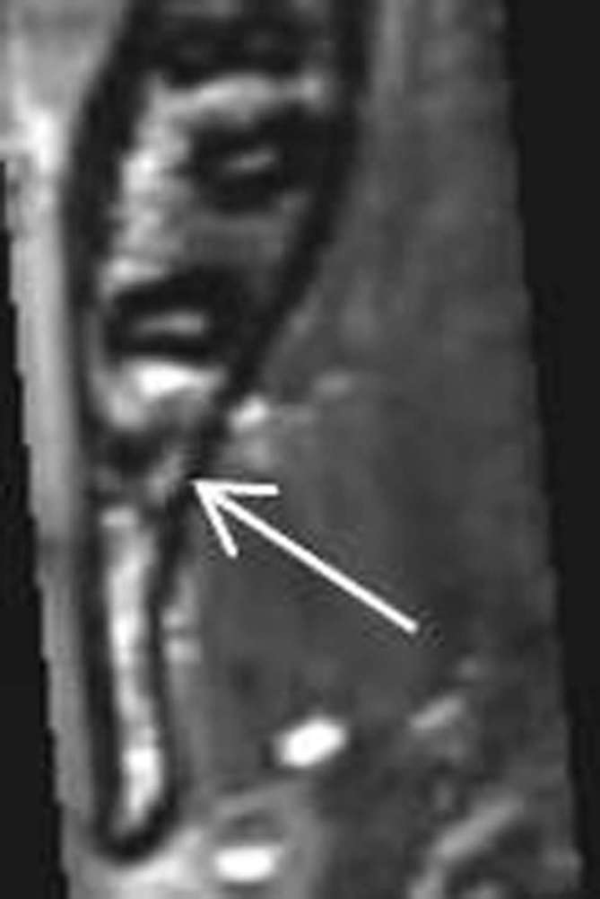
Axial MRI slice with inferior alveolar nerve at the lingual side of distal apex
Figure 2. .

Sagittal MRI slices at tooth 48 from buccal to lingual (left to right)
To collect comparable data for statistical evaluation in the current study, panoramic radiographs and MRIs were assessed according to the classification of Juodzbalys and Daugela.15 Assessment comprised six different parameters (M, R, A, C, B, S) describing the absolute position of the lower wisdom tooth and the relation to neighboring anatomical structures (Figure 4). Based on the collected data, the degree of expected surgical difficulty was graded from 0 to 4 for each parameter: conventional (0), simple (1), moderate (2), and complicated (3).The overall grade was determined by the highest grade among the 6 parameters.15 Parameter B was only assessed in MRI and not included in the comparison, because the two-dimensional panoramic radiographs did not show the bucco-lingual position of the third molar.
Figure 4. .

Grading according to Juodzbalys and Daugela 2013 (15)
Four investigators assessed the anonymized data sets twice, at intervals of at least one day. The investigators had different levels of experience and training and included two experienced oral surgeons with specialization in oral radiology (OS1 and OS2), one general dentist (G) and one general radiologist (R). All images were displayed on a standardized screen for radiological evaluation (Todoku, Monochrome LCD Monitor ME 253i2/C) in a quiet dark room; Sidexis radiological diagnostic software (Sirona Dental Systems, Inc. Dentsply Sirona, York, PA) was used for panoramic radiographs and SyngoMMWP VE31A (SiemensAG, Erlangen, Germany) for MRI. MRI assessment was performed on axial and sagittal layers. Image modification including magnification, contrast enhancing or brightness control of panoramic radiographs and MRIs, as well as movement through axial and sagittal MRI slices, was allowed according to individual preferences.
Statistical analysis was performed with SPSS software (IBM SPSS statistics 23.0, IBM Corporation, New York, NY). Agreement of panoramic radiography and MRI was evaluated and weighted κ was calculated to assess intra- and interobserver agreement (Figures 5 and 6).
Figure 5. .

Intraobserver agreement for panoramic radiography (PR) and MRI
Figure 6. .

Interobserver agreement for panoramic radiography (PR) and MRI
Results
All panoramic radiographs covered the full upper and lower jaws, showing dental and adjacent anatomical structures in two dimensions.
The MRI images showed the regions of the lateral teeth of the upper and lower jaws on both sides including neighboring anatomical structures. Image resolution in interpreted axial and sagittal reformations was highly detailed. It was possible to differentiate reliably between pulp and surrounding hard tooth tissue and bony structures. The dental follicle was displayed at the impacted third molars. The jaw bone could be well differentiated from the surrounding soft tissue. The inferior alveolar nerve and its dental branches were traceable, and even thin healthy sinus mucosa could be distinguished. Artefacts did not affect survey findings. There were no unexpected pathological or incidental findings.
Overall 56 lower third molars were investigated, with 448 assessments per method totaling 896 overall. In panoramic radiography, 2240 single parameters were evaluated and in MRI, 2688.
The first assessment run revealed the following overall staging results (Table 1):
Table 1.
Staging results of 1st assessment
| MRI 1 | Total | ||||
| 1 | 2 | 3 | |||
| PR 1 | 0 | 0 | 0 | 1 | 1 |
| 1 | 0 | 1 | 0 | 1 | |
| 2 | 2 | 10 | 4 | 16 | |
| 3 | 0 | 7 | 31 | 38 | |
| Total | 2 | 18 | 36 | 56 | |
PR (panoramic radiography)
Summing up results of all parameters in panoramic radiographs, 38 of 56 teeth (67%) were staged as “complicated” (Grade 3), 16 (28%) as “moderate” (Grade 2), one (2%) as “simple” (Grade 1), and one (2%) as conventional (Grade 0). In MRI only 36 teeth (64%) were evaluated as “complicated”, 18 (32%) as “moderate”, and two (3%) as “simple”. In 73% of cases, the two imaging techniques provided the same stagings. Weighted κa was 0.433.
In the second run (Table 2), 36 of 56 (64%) teeth were assessed as “complicated”, 19 (34%) were “moderate”, and one (2%) was “conventional” in panoramic radiographs. In MRI, 39 teeth (70%) were staged as “complicated”, 13 (23%) “moderate”, three (5%) “simple”, and one (2%) “conventional”. The second assessment showed a 77% agreement between panoramic radiography and MRI. Weighted κ was 0.495.
Table 2.
Staging results of 2nd assessment
| MRI 2 | Total | |||||
| 0 | 1 | 2 | 3 | |||
| PR 2 | 0 | 0 | 0 | 0 | 1 | 1 |
| 2 | 1 | 3 | 10 | 5 | 19 | |
| 3 | 0 | 0 | 3 | 33 | 36 | |
| Total | 1 | 3 | 13 | 39 | 56 | |
Detailed concordance of each parameter is as follows and is shown in Figure 7.
Figure 7. .

Percentual accordance PR versus MRI
Parameter M describes the position of the third molar relative to the second molar. Weighted κ was 0.606. Staging concurred in 72% of cases.
Parameter R refers to the position of the third molar in the ascending ramus. Weighted κ was 0.392. Consensus was reached in 57% of cases.
Parameter A concerns the vertical relationship between the third molar and the alveolar crest. Weighted κ was 0.568 and agreement was 60%.
Parameter C describes the position of the third molar in relation to the inferior alveolar nerve. Weighted κ was 0.142 and agreement was 56%.
Parameter S refers to the angulation of the third molar, measured by the angle between the longitudinal axis of third molar and the occlusal level of the first and second molars. Weighted κ was 0.597, with concordance of 78%
Parameter B was evaluated only in MRI. It refers to the bucco-lingual position, indicating greater difficulty with a closer position to the lingual wall. 28ht of 56 teeth (50%) were graded “complicated” (Grade 3), 19 (34%) “moderate” (Grade 2), 8 (14%) “simple” (Grade 1), and one (2%) “conventional” (Grade 0).
κ results for intra-observer agreement in panoramic radiography and MRI measurements are listed in Figure 3. κ results of investigator R (radiologist) were better for MRI, while the results of the other 3 investigators were better for panoramic views. (Figure 5)
Inter-observer agreement was evaluated between investigator OS1 (oral surgeon 1) and R (radiologist), G (general dentist) and OS2 (oral surgeon 2) and OS1 and OS2.
Investigators OS1 and OS2 show the best agreement, mainly seen in panoramic radiographs. Agreement was lowest between OS1 and R, especially in MRI. (Figure 6).
Discussion
To the best of our knowledge, this is the first study to address comparative wisdom tooth assessment in panoramic radiographs and MRIs. Findings suggest a positive answer to the question of whether dose-free dental MRI is a potential alternative for advanced preoperative radiological assessment of lower impacted lower third molars. MRI provided images with quite good resolution that displayed all relevant anatomical structures and so supplied acceptable quality for diagnostic pre-surgical third molar assessment.
Standardized assessment of lower third molars followed a recently published classification by Juodzbalys and Daugela15. This classification system is based on 75 literature sources, taking traditional radiological classifications into account. It is the only classification so far that allows three-dimensional assessment of third molar position in the jaw, and so is uniquely suitable for interpreting panoramic radiographs and cross-sectional images.15
Staging results for panoramic radiography and MRI concurred in 73% after the first run (Table 1) and in 77% after the second run (Table 2), indicating in general similar grading independent of radiological method. Moreover, MRI as a cross-sectional technique, provides the advantage of three-dimensional assessment of relevant anatomical details.
The vertical position of the wisdom tooth to the second molar and the alveolar crest, as well as angulation, seems to be well displayed in both modalities. This resulted in a high accordance in parameters M, A, and S. On the other hand, parameters C and R, reflecting position in relation to the inferior alveolar nerve and in the ascending ramus, showed greater differences. The assessment of the inferior alveolar nerve (parameter C) is superior in MRI, as it displays the exact position in the bucco-lingual and cranio-caudal directions when superimposition is ruled out.16, 17 Likewise, the position in relation to the ascending ramus (parameter R) cannot be sufficiently assessed in panoramic radiography due to overlapping. The definition of the parameter itself, which allows a wide range of interpretation and the chosen MRI layer, may explain the weak agreement. Finally, there is parameter B, reflecting the bucco-lingual location in the mandible, which is only assessable in MRI. Here, it should be noted that the preoperative awareness of the three-dimensional tooth position can shorten and facilitate the surgery.18, 19
The two oral surgeons achieved the highest κ results for inter-observer-agreement, very likely because of their specialization in oral radiology with similar levels of training and long experience (Figure 6). Regarding agreement on the first and second runs, 3 of 4 investigators were more consistent in panoramic radiography than in MRI (Figure 5). This could be because the diverging investigator, a radiologist, was more used to MRI data sets, while the other three examiners, two oral surgeons and one general dentist, were more accustomed to interpreting panoramic radiographs. Clearly, the interpretation of MRI data involves a learning curve.
Even though conventional X-ray imaging techniques are well established in dentistry, it must be borne in mind that lifetime cancer risk increases with the ionizing radiation from every single scan, especially in radiation sensitive adolescents.10 This should be considered seriously if in the light of existing research, MRI could be proposed as a dose-free diagnostic alternative.16,20–24 To date, poor image quality and low spatial resolution compared to CBCT and CT are some reasons for the reluctance to use of MRI in dentistry, but those deficits are being met with growing field intensity and individualized coils for small detail accuracy.12 A MRI system with least 3.0 tesla, as used here, is certainly preferable for dental MRI.25 There already are reports of substantial improvements in dental MRI when intraoral coils produce high resolution images with short acquisition times. However, these innovations are not as yet in widespread use.26, 27
Artefacts due to dental restorations may be a shortcoming of MRI in dentistry as they diminish the diagnostic value of the images. Dental materials may distort the applied radio frequency field or the static magnetic field and affected areas appear more or less hypointense because of signal loss.28, 29 As the literature and the present study demonstrate, MRI can nonetheless be satisfactory for radiological dental evaluation,12, 14,20,23 despite some disadvantages that are regularly cited that speak against its routine use in dentistry. These include the current acquisition times of more than 20 min, high costs and relatively low accessibility, as well as the discomfort to the patient of being a fixed position in a noisy machine for an extended period.30, 31
Conclusion
The presented study demonstrates that MRI not only provides much the same information that panoramic radiography usually does, but also has the advantages of a dose-free three-dimensional view. This may facilitate and shorten third molar surgery. Image interpretation, however, can differ depending on training and experience.
REFERENCES
- 1. de Carvalho RW, de Araújo Filho RC, do Egito Vasconcelos BC. Assessment of factors associated with surgical difficulty during removal of impacted maxillary third molars. J Oral Maxillofac Surg 2013; 71: 839–45. doi: 10.1016/j.joms.2013.01.001 [DOI] [PubMed] [Google Scholar]
- 2. Steed MB. The indications for third-molar extractions. J Am Dent Assoc 2014; 145: 570–3. doi: https://doi.org/10.14219/jada.2014.18 [DOI] [PubMed] [Google Scholar]
- 3. Gbotolorun OM, Arotiba GT, Ladeinde AL. Assessment of factors associated with surgical difficulty in impacted mandibular third molar extraction. J Oral Maxillofac Surg 2007; 65: 1977–83. doi: 10.1016/j.joms.2006.11.030 [DOI] [PubMed] [Google Scholar]
- 4. Cassetta M, Pranno N, Barchetti F, Sorrentino V, Lo Mele L. 3.0 Tesla MRI in the early evaluation of inferior alveolar nerve neurological complications after mandibular third molar extraction: a prospective study. Dentomaxillofac Radiol 2014; 43: 20140152. doi: 10.1259/dmfr.20140152 [DOI] [PMC free article] [PubMed] [Google Scholar]
- 5. Roeder F, Wachtlin D, Schulze R. Necessity of 3D visualization for the removal of lower wisdom teeth: required sample size to prove non-inferiority of panoramic radiography compared to CBCT. Clin Oral Investig 2012; 16: 699–706. doi: 10.1007/s00784-011-0553-8 [DOI] [PubMed] [Google Scholar]
- 6. De Vos W, Casselman J, Swennen GR. Cone-beam computerized tomography (CBCT) imaging of the oral and maxillofacial region: a systematic review of the literature. Int J Oral Maxillofac Surg 2009; 38: 609–25. doi: 10.1016/j.ijom.2009.02.028 [DOI] [PubMed] [Google Scholar]
- 7. Horner K, O'Malley L, Taylor K, Glenny AM. Guidelines for clinical use of CBCT: a review. Dentomaxillofac Radiol 2015; 44: 20140225. doi: 10.1259/dmfr.20140225 [DOI] [PMC free article] [PubMed] [Google Scholar]
- 8. Ludlow JB, Timothy R, Walker C, Hunter R, Benavides E, Samuelson DB, et al. Effective dose of dental CBCT-a meta analysis of published data and additional data for nine CBCT units. Dentomaxillofac Radiol 2015; 44: 20140197. doi: 10.1259/dmfr.20140197 [DOI] [PMC free article] [PubMed] [Google Scholar]
- 9. Schulze D, Heiland M, Thurmann H, Adam G. Radiation exposure during midfacial imaging using 4- and 16-slice computed tomography, cone beam computed tomography systems and conventional radiography. Dentomaxillofac Radiol 2004; 33: 83–6. doi: 10.1259/dmfr/28403350 [DOI] [PubMed] [Google Scholar]
- 10. Smith-Bindman R, Lipson J, Marcus R, Kim KP, Mahesh M, Gould R, et al. Radiation dose associated with common computed tomography examinations and the associated lifetime attributable risk of cancer. Arch Intern Med 2009; 169: 2078–86. doi: 10.1001/archinternmed.2009.427 [DOI] [PMC free article] [PubMed] [Google Scholar]
- 11. Konouchi H, Yanagi Y, Hisatomi M, Matsuzaki H, Takenobu T, Unetsubo T, et al. MR imaging diagnostic protocol for unilocular lesions of the jaw. Jpn Dent Sci Rev 2012; 48: 81–91. doi: 10.1016/j.jdsr.2012.01.001 [DOI] [Google Scholar]
- 12. Tymofiyeva O, Rottner K, Jakob PM, Richter EJ, Proff P. Three-dimensional localization of impacted teeth using magnetic resonance imaging. Clin Oral Investig 2010; 14: 169–76. doi: 10.1007/s00784-009-0277-1 [DOI] [PubMed] [Google Scholar]
- 13. Gray CF, Redpath TW, Smith FW, Staff RT. Advanced imaging: magnetic resonance imaging in implant dentistry. Clin Oral Implants Res 2003; 14: 18–27. doi: 10.1034/j.1600-0501.2003.140103.x [DOI] [PubMed] [Google Scholar]
- 14. Baumann P, Widek T, Merkens H, Boldt J, Petrovic A, Urschler M, et al. Dental age estimation of living persons: Comparison of MRI with OPG. Forensic Sci Int 2015; 253: 76–80. doi: 10.1016/j.forsciint.2015.06.001 [DOI] [PubMed] [Google Scholar]
- 15. Juodzbalys G, Daugela P. Mandibular third molar impaction: review of literature and a proposal of a classification. J Oral Maxillofac Res 2013; 4: e1. doi: 10.5037/jomr.2013.4201 [DOI] [PMC free article] [PubMed] [Google Scholar]
- 16. Gahleitner A, Watzek G, Imhof H. Dental CT: imaging technique, anatomy, and pathologic conditions of the jaws. Eur Radiol 2003; 13: 366–76. doi: 10.1007/s00330-002-1373-7 [DOI] [PubMed] [Google Scholar]
- 17. Peker I, Sarikir C, Alkurt MT, Zor ZF. Panoramic radiography and cone-beam computed tomography findings in preoperative examination of impacted mandibular third molars. BMC Oral Health 2014; 14: 71–8. doi: 10.1186/1472-6831-14-71 [DOI] [PMC free article] [PubMed] [Google Scholar]
- 18. Matzen LH, Wenzel A. Efficacy of CBCT for assessment of impacted mandibular third molars: a review - based on a hierarchical model of evidence. Dentomaxillofac Radiol 2015; 44: 20140189. doi: 10.1259/dmfr.20140189 [DOI] [PMC free article] [PubMed] [Google Scholar]
- 19. Susarla SM, Dodson TB. Preoperative computed tomography imaging in the management of impacted mandibular third molars. J Oral Maxillofac Surg 2007; 65: 83–8. doi: 10.1016/j.joms.2005.10.052 [DOI] [PubMed] [Google Scholar]
- 20. Fiehler J. Possibilities and perspectives in dental MRI. Vol. 65: The British Institute of Radiology.; 2014. 741–9. [Google Scholar]
- 21. Chau A. Comparison between the use of magnetic resonance imaging and conebeam computed tomography for mandibular nerve identification. Clin Oral Implants Res 2012; 23: 253–6. doi: 10.1111/j.1600-0501.2011.02188.x [DOI] [PubMed] [Google Scholar]
- 22. Eggers G, Rieker M, Fiebach J, Kress B, Dickhaus H, Hassfeld S. Geometric accuracy of magnetic resonance imaging of the mandibular nerve. Dentomaxillofac Radiol 2005; 34: 285–91. doi: 10.1259/dmfr/89236515 [DOI] [PubMed] [Google Scholar]
- 23. Ferretti F, Malventi M, Malasoma R. Dental magnetic resonance imaging: study of impacted mandibular third molars. Dentomaxillofac Radiol 2009; 38: 387–92. doi: 10.1259/dmfr/29929241 [DOI] [PubMed] [Google Scholar]
- 24. Imamura H, Sato H, Matsuura T, Ishikawa M, Zeze R. A comparative study of computed tomography and magnetic resonance imaging for the detection of mandibular canals and cross-sectional areas in diagnosis prior to dental implant treatment. Clin Implant Dent Relat Res 2004; 6: 75–81. doi: 10.1111/j.1708-8208.2004.tb00029.x [DOI] [PubMed] [Google Scholar]
- 25. Fischbach F, Müller M, Bruhn H. Magnetic resonance imaging of the cranial nerves in the posterior fossa: a comparative study of t2-weighted spin-echo sequences at 1.5 and 3.0 tesla. Acta Radiol 2008; 49: 358–63. doi: 10.1080/02841850701824127 [DOI] [PubMed] [Google Scholar]
- 26. Ludwig U, Eisenbeiss A-K, Scheifele C, Nelson K, Bock M, Hennig J, et al. Dental MRI using wireless intraoral coils. Sci Rep 2016; 6: 1–11. doi: 10.1038/srep23301 [DOI] [PMC free article] [PubMed] [Google Scholar]
- 27. Idiyatullin D, Corum C, Moeller S, Prasad HS, Garwood M, Nixdorf DR. Dental magnetic resonance imaging: making the invisible visible. J Endod 2011; 37: 745–52. doi: 10.1016/j.joen.2011.02.022 [DOI] [PMC free article] [PubMed] [Google Scholar]
- 28. Tymofiyeva O, Vaegler S, Rottner K, Boldt J, Hopfgartner AJ, Proff PC, et al. Influence of dental materials on dental MRI. Dentomaxillofac Radiol 2013; 42: 20120271. doi: 10.1259/dmfr.20120271 [DOI] [PMC free article] [PubMed] [Google Scholar]
- 29. Klinke T, Daboul A, Maron J, Gredes T, Puls R, Jaghsi A, et al. Artifacts in magnetic resonance imaging and computed tomography caused by dental materials. PLoS One 2012; 7: e31766. doi: 10.1371/journal.pone.0031766 [DOI] [PMC free article] [PubMed] [Google Scholar]
- 30. Chou IJ, Tench CR, Gowland P, Jaspan T, Dineen RA, Evangelou N, et al. Subjective discomfort in children receiving 3 T MRI and experienced adults’ perspective on children’s tolerability of 7 T: a cross-sectional questionnaire survey. BMJ Open 2014; 4: e006094. doi: 10.1136/bmjopen-2014-006094 [DOI] [PMC free article] [PubMed] [Google Scholar]
- 31. Okada T, Nakai T. Silent fMRI acquisition methods for large acoustic noise during scan. Magn Reson Med Sci 2003; 2: 181–7. doi: 10.2463/mrms.2.181 [DOI] [PubMed] [Google Scholar]