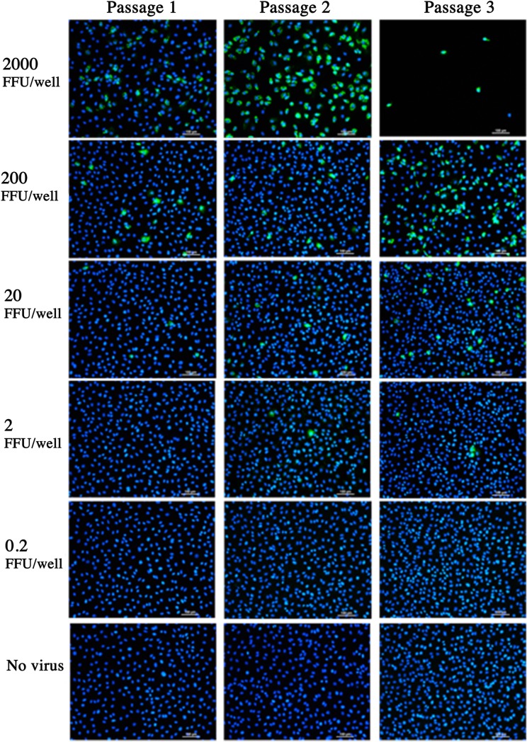
Fig 3. The ability to detect very low FFU of live RV.
Monolayers of MA104 cells were incubated for 24h with 0.2 to 2000 FFU live RV. Uninfected MA104 cells were used as a negative control. Culture supernatant was used to infect new MA104 monolayers, and previously infected monolayers were visualised by FFA. DAPI (blue) indicates cell nuclei and FITC (green) indicates RV infection. Stained monolayers were visualised using Nikon TiE inverted fluorescent microscope. Scale bar = 100μm. Images representative of 3 replicates per MOI for each passage.

