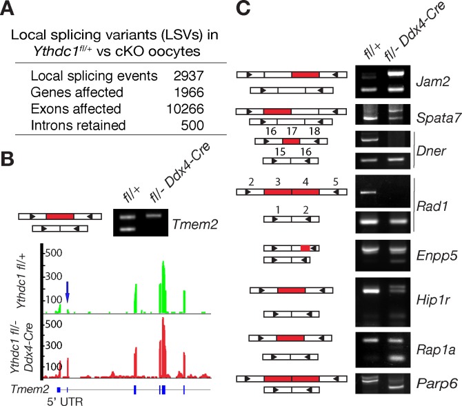
Fig 5. Changes in the splicing landscape in Ythdc1-deficient oocytes.
Oocytes were collected from 6-week-old Ythdc1fl/+ and Ythdc1fl/- Ddx4-Cre females. (A) Summary of local splicing variants (LSVs) identified by MAJIQ. Significant LSVs: ΔPSI (difference in percentage spliced in) > 0.2 and q < 0.05. (B) PCR validation and gene track view of one exon-skipping LSV in Tmem2. (C) PCR validation of LSVs affecting internal exons. Exons are represented as rectangles but not in scale. Skipped or retained exons are shown in red. Triangles denote the positions of PCR primers. Each PCR assays was performed three times using different samples.

