
Figure 1. Interactions that regulate T cell responses.
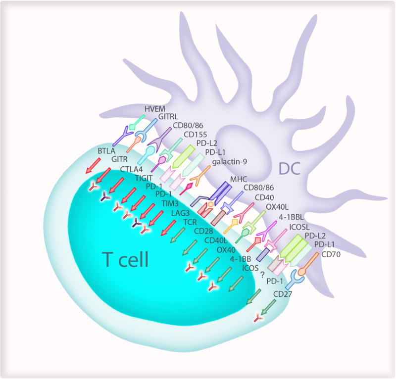
Antigen presenting cells such as dendritic cells (DCs) regulate T cell response to specific pathogens or antigens from malignant cells. The T cell receptors (TCR) on antigen-specific T cells first recognise their cognate antigen via the major histocompatibility complex (MHC) molecules on antigen presenting cells. This step has to be followed by signals to CD28 on T cells from CD80 on the APC and is described as “signal 2”. Several different ligands on DCs then provide signals to T cells which decide the quality and duration of the effector response (green arrows). These include CD40/CD40 ligand (CD40L); OX40/OX40 ligand (OX40L); 4-1BB (CD137)/4-1BB ligand (41BBL; CD137 Ligand); ICOS (Inducible T-cell COStimulator; CD278)/ICOS Ligand (ICOS-L); CD27/CD70. There are also signals to suppress immune responses (red arrows) to maintain self tolerance and limit the duration of immune responses to minimize bystander damage to host tissue. These include LAG3 (lymphocyte activation gene 3); MHC class II; TIM3 (T cell immunoglobulin and mucin-domain containing-3; HAVCR2 in humans)/galectin-9; PD-1 (programmed cell death-1)/PD-L1 (programmed cell death-1-ligand 1) and PD-L2 (programmed cell death-1-ligand 2); TIGIT (T cell immunoreceptor with Ig and ITIM domains)/CD155; CTLA4 (cytotoxic T-lymphocyte-associated protein 4)/CD86 or CD80; GITR (Glucocorticoid-induced TNFR-related protein)/GITR-L (GITR-ligand) and BTLA (B and T lymphocyte attenuator)/HVEM (Herpesvirus entry mediator). Antibody symbol represents pathways being tested in current clinical trials. The “?” refers to an unknown receptor which “activates” T cells. The “red” antibodies indicate pathways undergoing clinical trials for cancer and the “dark coloured” antibodies indicate clinical use.