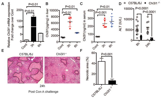
Fig. 1. Chi3l1 contributes to Con A-induced liver injury in mice.
Hepatic mRNA expression of Chi3l1 (A), and protein levels of Chi3l1 in liver (B) and serum (C) were measured in male WT (C57BL/6J) mice treated with 15 mg/kg Con A by intravenous injection (n=3–5 mice per group). (D) Serum ALT levels were determined at 8 and 24 h post-Con A treatment (n=15–22 mice per group). (E) Shown are representative H&E-stained liver sections from at least 4 mice per group at 24h after Con A treatment (magnification, 100×). Necrotic areas are outlined. (F) Necrotic areas are quantified by Image J. P values are as indicated. Two-tailed, unpaired t test in D and F; one-way ANOVA in A, B, C.

