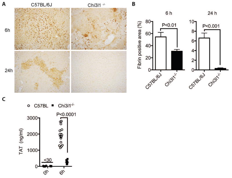
Fig. 3. Chi3l1−/− mice exhibit attenuated intrahepatic coagulation upon Con A challenge.
(A–B) Male WT and Chi3l1−/− were treated with Con A as described in Fig. 1 for 6h and 24h. Immunohistochemical staining for fibrin(ogen) (magnification: 200x) were performed and quantified. Images shown are representative of at least 4 mice per group. (C) The levels of TAT complex were measured in plasma of WT and Chi3l1−/− mice at 6h post-Con A treatment (n=14–15 per group). P values are as indicated. Two- tailed, unpaired t test were performed in B, D–F.

