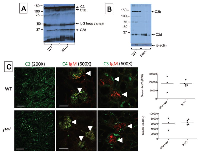
Figure 2. Complement activation in the kidneys of mice with ischemic acute kidney injury.

Wild-type (WT; C57BL/6) and mice with heterozygous deficiency of factor H (fH+/−) were subjected to ischemia/reperfusion of the kidneys. After 24 hours serum and tissues were collected. A) Western blot analysis of plasma from wild-type and factor H deficient mice showing levels of circulating intact C3. C3 fragments were detected using a monoclonal antibody to C3d (mAB 3d11) B) Western blot analysis of kidney lysates for C3b in factor H deficient mice with ischemic acute kidney injury compared to wild-type controls. The blot was re-probed with an antibody for β-actin as a loading control. C) Tissue sections were stained using antibodies to murine C3, C4, and IgM. Immunofluorescence microscopy for C3, C4, and IgM revealed C3 deposits throughout the tubulointerstitium of both strains of mice. High-powered views of the glomeruli (indicated with arrowheads) demonstrates the deposits of IgM and C4 co-localize within the glomeruli of both strains of mice. unpaired student t test showed no significant difference in both genotypes. Scale bar = 100 μm for 200X images and 50 μm for 600X images. Fluorescence data shown is representative of two experiments from which 3–5 mice from each group were analyzed. Each data point represents one mouse, and the bar represents the group mean.
