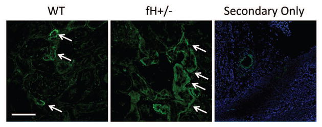
Figure 3. C6 deposition in the kidneys of mice with ischemic acute kidney injury.

Wild-type (WT; C57BL/6) and mice with heterozygous deficiency of factor H (fH+/−) were subjected to ischemia/reperfusion of the kidneys. After 24 hours tissues were collected, and kidney sections from four mice in each group were stained for C6.. Original magnification x600 and scale bar = 50 μm for the WT and fH+/− panels. Original magnification x200 and scale bar = 100 μm for the secondary only panel. Image shown is representative of four mice that were analyzed from two independent experiments.
