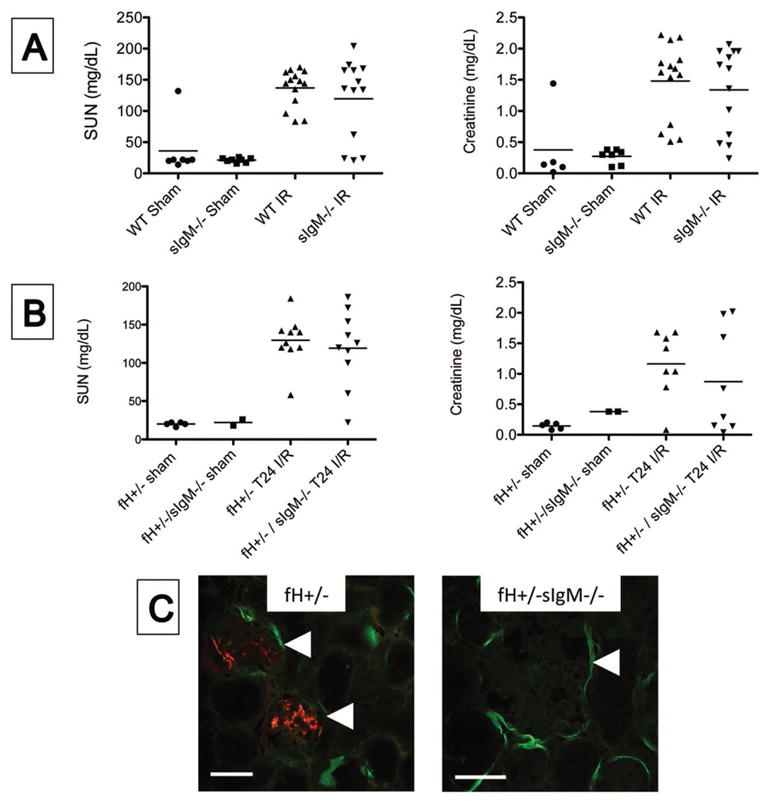
Figure 5. IgM does not contribute to kidney injury after ischemia/reperfusion.

Wild-type (WT) and mice that cannot generate soluble IgM (sIgM−/−) were subjected to ischemia/reperfusion of the kidneys. A) After 24 hours the serum urea nitrogen (SUN) and serum creatinine levels were measured using a NADH oxidation assay and a picric acid chromogenic assay, respectively. There was not a significant difference between WT and sIgM−/− mice when compared by ANOVA with Tukey post test. B) Factor H deficient (fH+/− mice) and mice deficient in both factor H and soluble IgM (fH+/−sIgM−/− mice) were subjected to ischemia/reperfusion of the kidneys. After 24 hours the SUN and serum creatinine levels were using the same assays as in A. There was not a significant difference between the two strains when compared using ANOVA with Tukey post test. C) Tissue sections were stained with antibodies to murine C3 and IgM. Immunofluorescence microscopy for C3 (green) and IgM (red) revealed IgM deposits in the glomeruli of fH+/− mice but not of fH+/−sIgM−/− mice. Original magnification 200X for fH+/− and 600X for fH+/−sIgM−/−. Glomeruli are indicated with arrowheads. Scale bar = 100 μm for 200X and 50 μm for 600X. Data shown A is from three ischemia experiments with 6–14 mice per experiment, and data in B is from four experiments 5–9 mice per experiment. Each mouse is represented by a data-point, and the mean for each group is shown. Images in C are representative of three mice from two experiments.
