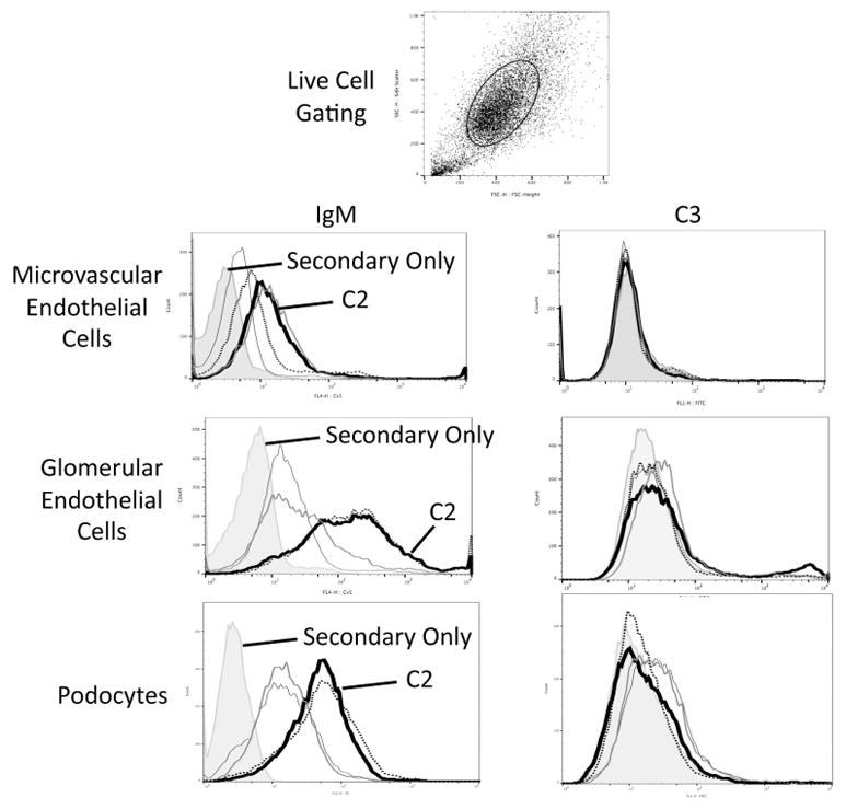
Figure 6. Natural IgM binds to glomerular cells in vitro.

Murine microvascular endothelial cells, glomerular endothelial cells, and podocyte cell lines were grown in culture and were incubated with several different natural IgM clones. Binding of IgM to live cells was examined by flow cytometry using an allophycocyanin (APC)-conjugated antibody to mouse IgM (μ chain). The gating strategy to examine live cells is shown. Several of the IgM clones bound to cells. Secondary antibody only staining is shown with the shaded graphs. One of the monoclonal IgM antibodies, C2, bound to all three cell types and is represented with a dark black line. In a separate set of experiments the cells were incubated with purified IgM and serum from IgM deficient mice, and C3 deposition on the cells was measured by flow cytometry using a fluorescein (FITC)-conjugated antibody to C3. Each experiment was performed at least three times independently, and representative results are shown.
