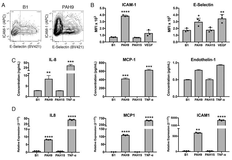
Figure 5. Induction of pro-inflammatory and pro-adhesive mediators in HUVECs by recombinant IPAH plasmablast mAbs.

HUVEC cells were stimulated with IPAH mAbs, isotype control mAb, or positive control cytokines. Cells were harvested after 6 hours and stained for ICAM-1 and E-selectin. Additional cultures were harvested after 24 hours for qPCR and cell supernatant ELISAs. Negative control antibody B1 was derived from sequencing the plasmablasts of a patient with bacterial infection. (A) Representative flow cytometric staining of mAb-stimulated HUVECs. Full gating strategy is displayed in Supplemental Figure S5. (B) Mean fluorescence intensity of ICAM-1 and E-selectin staining on HUVECS 8 hours following addition of the mAb. Data shown is representative of two independent experiments with three samples per experiment. (C) ELISA measurement of MCP-1, IL-8, and endothelin-1 secreted by mAb-stimulated HUVECs. (D) qPCR gene expression analysis of HUVECS following overnight stimulation. For panels (B-D), Each value represents the mean ± SD of three replicate samples per experiment, with similar results confirmed by at least two independent experiments. ***P<0.001, **P<0.01, *P<0.05 by one-way ANOVA with Tukey's post-hoc test.
