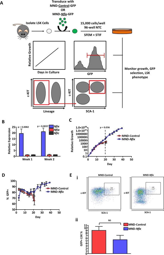
Figure 1. Nfix induces longevity in HSPC ex vivo culture.

(A) Experimental schematic. 48 hours post-transduction, LSK cells transduced with MND-Control or MND-Nfix were re-plated in 96-well non-tissue culture treated plates in serum-free expansion medium (SFEM) supplemented with mSCF, mTPO, mIGF-2, and hFGF-a (STIF). Every 48-72 hours of culture, cells were counted, assessed for GFP+ cells, and passaged 1:4. Cells were also periodically assessed for LSK immuno-phenotype. (B) Relative expression of NFI-family genes in NFIX+ cells compared to control cells, quantified by qRT-PCR (n = 3). Tbp was used as a housekeeping gene. (C) Relative growth of control and NFIX+ cells during ex vivo culture (n = 4). Dotted line indicates the divergence in relative growth between control and NFIX+ cells. (D) GFP percentage of control and NFIX+ cells during ex vivo culture, assessed by flow cytometry (n = 4). (E) Percentage of control and NFIX+ cells with an LSK immuno-phenotype at day seven of ex vivo culture, depicted as a (i) representative dot plot and (ii) bar plot (n = 6). All values represent mean ± standard deviation. NS denotes not significant.
