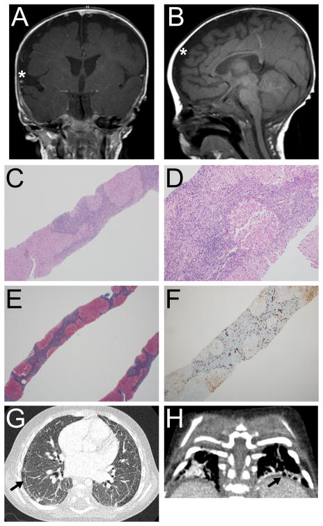
Figure 1.
Head MRI, liver pathology, and chest CT of the proband. A and B: Coronal and sagittal T1-weighted head MRI images obtained at 5 months corrected age showing incomplete closure of the Sylvian fissures (asterisk in A), and bilateral frontoparietal cerebral volume loss (asterisk in B). C–F: Liver pathology. Architectural distortion (4X) with nodular formation (C); portal chronic inflammation with focal steatosis (10X) (D); Masson trichrome stain (2X) with well-developed cirrhosis and bridging fibrosis (E); and CK7 immunostain (4X) showing extensive bile ductular reaction (F). G and H: Chest CT (18 months) showing subpleural cystic changes (arrows) and interstitial disease.

