
Figure 2.
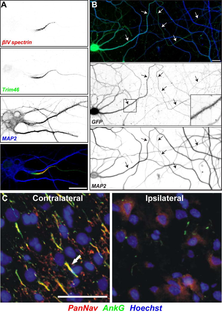
Axon initial segments in health and disease. (A) A cultured hippocampal neuron immunolabeled using antibodies against βIV spectrin (red), Trim46 (green), and MAP2 (blue). Scalebar = 10 μm. (B) A cultured hippocampal neuron infected with virus to express GFP and ankG shRNA. The neuron has lost neuronal polarity; the former axon (arrows) acquires dendritic features including MAP2 and spines (box and inset). Scalebar = 20 μm. Modified from Hedstrom et al.2 (C) Na+ channels are lost from the AIS following ischemic injury. Contralateral (left) and ipsilateral (right) regions of rat cortex were immunolabelled for Na+ channels (PanNav, red), ankG (green), and Hoechst to label nuclei (blue). The sections shown are from a brain collected 24 hr after MCAO. Arrows indicate labeled AIS in layer 2/3 cortex. Scale bar = 50 μm. Modified from Schafer et al.85