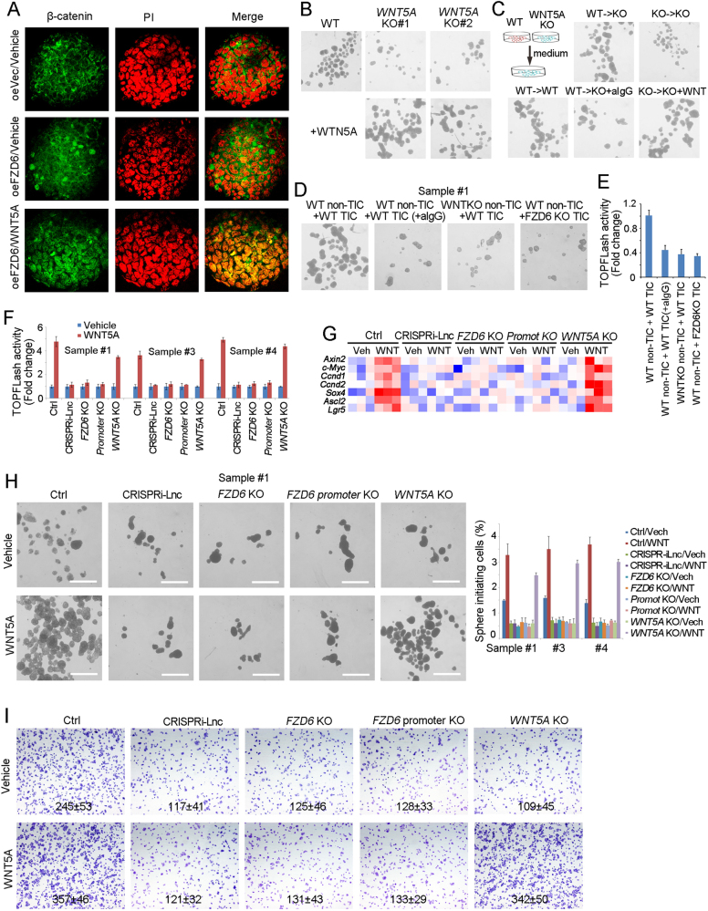
Fig. 6.
WNT5A activates Wnt/β-catenin signaling through FZD6-dependent manner. a The indicated treated spheres were examined for β-catenin nuclear translocation by confocal microscope. b Sphere formation assays were performed using WNT5A knockout cells, supplemented with recombinant WNT5A protein or not. c WT and WNT5A knockout cells were used for 1 week’s sphere formation and the supernatant medium were obtained for sphere formation of WNT5A knockout cells. Two weeks later, the typical spheres were shown. d, e WT and WNT5A KO non-TICs were sorted and mixed with TICs at ratio of 1:1, followed by oncosphere formation assay d or TOPFLash assay e. aIgG, WNT5A antibody IgG. f The indicated cells were treated with WNT5A, and Wnt/β-catenin activation was examined using TOPFLash methods. g The indicated cells were treated with WNT5A for 2 days, and the expression levels of Wnt/β-catenin target genes were examined by real-time PCR. The expression data were shown as heatmap. h In all, 1000 indicated cells were incubated in sphere formation medium with/o WNT5A for 2 weeks. Typical pictures were shown in left panels and calculated sphere-initiating ratios were shown in right panels. Scale bars, 500 μm. i The indicated cells were incubated with WNT5A, and tumor invasion capacity was observed with transwell assay. Typical images and invasive cell numbers (mean ± s.d.) were shown. Data were shown as means ± s.d. *P < 0.05 by two-tailed Student’s t-test. Data are representative of three independent experiments

