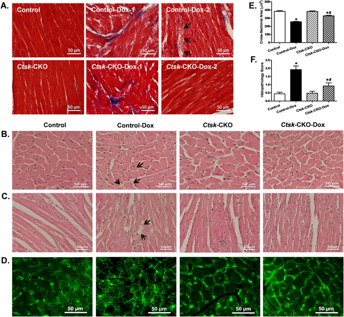
Fig. 5. Histological analyses in hearts from control and cardiac-specific Ctsk-CKO mice treated with or without doxorubicin.
a Representative images of Masson trichrome staining for fibrosis (×400; scale bar = 50 μm); b Representative H&E staining micrographs of transverse sections of left ventricular myocardium (×400; scale bar = 50 μm); c Representative H&E staining micrographs of longitudinal sections of left ventricular myocardium (×200; scale bar = 50 μm); d Representative Lectin staining of transverse sections of left ventricular myocardium (×400; scale bar = 50 μm); e Quantitative cardiomyocyte cross-sectional (transverse) area using measurements of 230 cardiomyocytes from three mice per group; f Semi-quantitative vacuolization and myofibrillar degeneration of the cardiomyocytes by scoring scales from 25 slides per group. Mean ± SEM, *p < 0.05 vs. Control group, #p < 0.05 vs. Control-Dox group. Arrow: cytoplasmic vacuolization and myofibrillar degeneration

