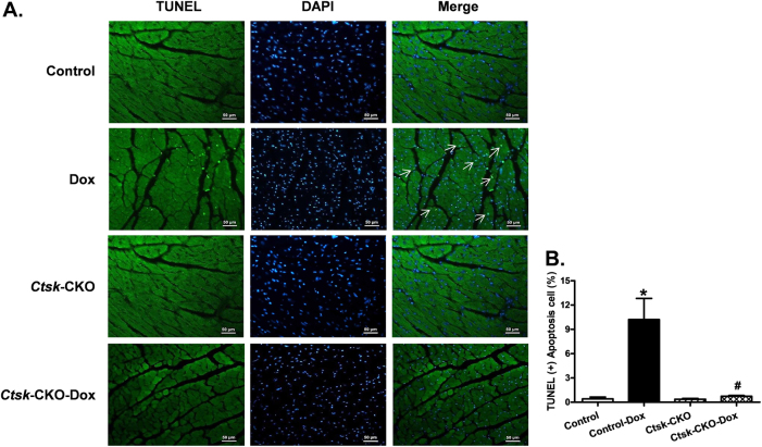
Fig. 8. TUNEL staining of apoptosis in myocardium from control and cardiac-specific Ctsk-CKO mice treated with or without doxorubicin.
a Representative photomicrograph of the TUNEL staining; b Quantitative analysis of the apoptotic cells. Mean ± SEM, *p < 0.05 vs. Control group, #p < 0.05 vs. Control-Dox group. TUNEL-positive nuclei were visualized with green fluorescein. All nuclei were stained with DAPI shown in blue color. Apoptotic cells are indicated by white arrows

