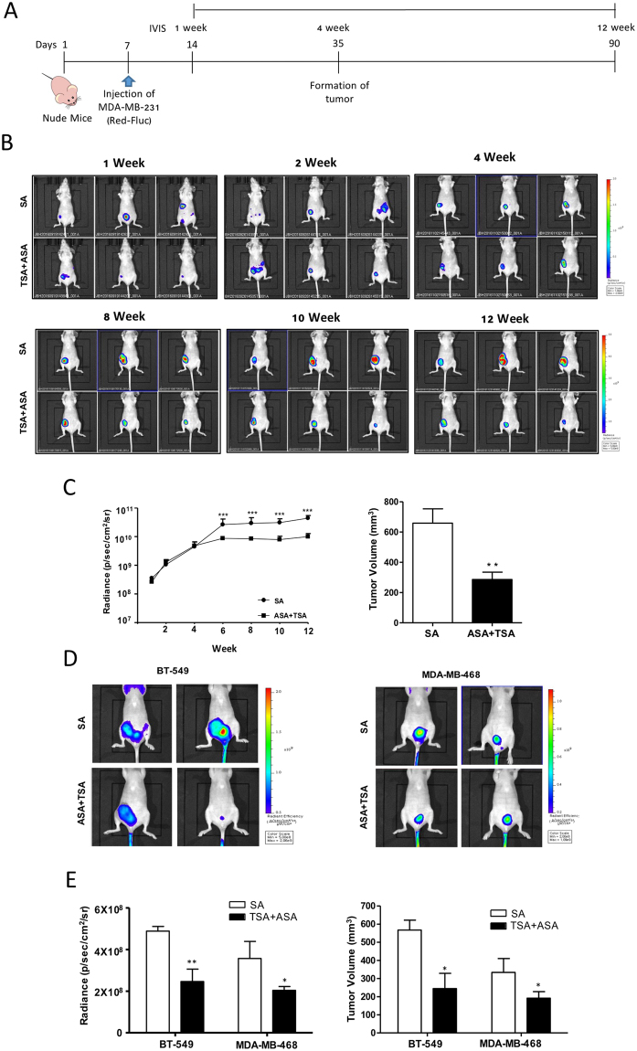
Figure 7.
Hyperacetylation significantly reduces tumor growth in vivo. (A–C) Schematic representation of the treatment regime for the administration of 20 mg/(kg·day) ASA plus subcutaneous injection of 0.5 mg/kg TSA, or SA (as a negative control) three times weekly in MDA-MB-231 cells xenografts. Female nude mice were injected with 1.5 × 10⁶ MDA-MB-231-FLuc cells. After the formation of detectable tumors according to IVIS imaging (7 days post injection), mice were weighed and randomly assigned to SA or ASA + TSA groups (n = 7, animals/group) (A). Representative IVIS images obtained after luciferin injection at different days post-injection of MDA-MB-231-FLuc cells, showing reduced tumor growth in hyperacetylated mice (B). Tumor-growth curves and tumor weights for both animal groups are shown for up to 90 days. Based on a slope comparison test, the treated animals showed a marked reduction in tumor growth [t (6) = 2.6]. ***P < 0.001 versus SA-treated control mice by an unpaired t-test (C). (D and E) Female nude mice were injected with 1.5 × 10⁶ BT-549 or MDA-MB-468 cells. After the formation of detectable tumors according to IVIS imaging with MMPSense750, the mice were weighed and randomly assigned to SA or ASA + TSA group (n = 4 animals/group). Tumor growth and tumor volume in both animal groups were assessed for up to 60 days. Columns represent the mean (n = 4); bars represent the SE. *P < 0.05; **P < 0.01 versus SA-treated control mice as determined by an unpaired t-test.

