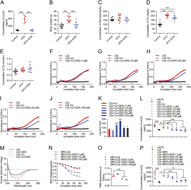
Fig. 2. DDS decreases the level of oxLDL.
a Blood serum oxLDL content was detected by oxLDL ELISA kit in control, HFD and DDS treated HFD groups. b Body weight, c serum glucose, d TC, e TG were measured in previous groups. The results were represented as mean ± SEM, n = 6–10 animals per group. The groups were compared by one-way ANOVA and followed by Dunnett’s multiple comparison tests. Differences were considered significant at ***p < 0.001. f-j Measurement of the kinetics of oxidation of LDL by continuously monitoring the change of UV absorbance at 234 nm; the UV absorbance curve of different groups: native LDL treatment with BHT, LDL-oxidized modification by Cu2+ and the treatment with dose-dependent DDS (1 µM, 10 µM, 25 µM, 50 µM, 100 µM). These three treatments were mimicked by the Boltzman sigmoidal nonlinear regression model. k The lag time of LDL oxidation in each groups. l oxLDL content by an oxLDL ELISA kit. The results were represented as mean ± SEM of four independent experiments. The treatment groups were compared by one-way ANOVA and followed by Dunnett’s multiple comparison tests. Differences were considered significant at *p < 0.05, **p < 0.01, ***p < 0.001. m CD measurement of control group, LDL treated with Cu2+ group, LDL oxidized with Cu2+ pre-treatment with DDS group. n MPO residual activity detected with absorbance at 670 nm. o The statistical analysis of MPO peroxidase activity. p oxLDL was detected using an ELISA kit. Mean ± SEM of three independent experiments. The treatment groups were compared by one-way ANOVA and followed by Dunnett’s multiple comparison tests. Differences were considered significant at *p < 0.05, **p < 0.01, ***p < 0.001

