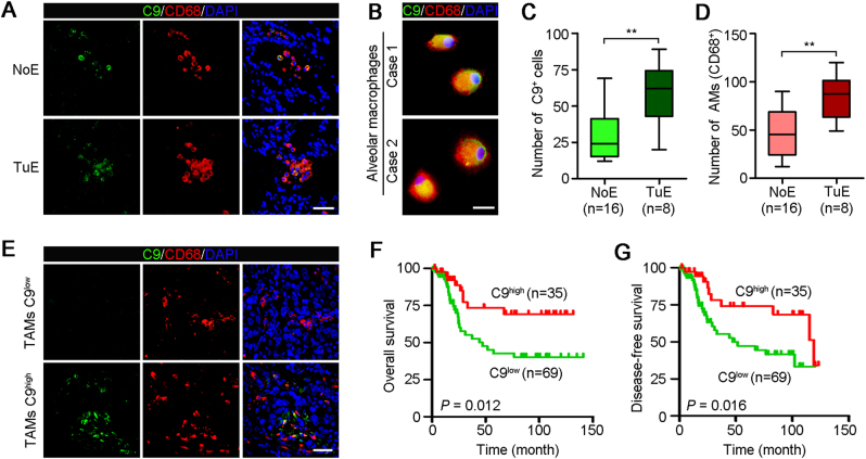
Fig. 3. TAMs high expression of C9 associates with superior survival for NSCLC patients.
a Representative images of two NSCLC adjacent nontumor tissues with no effect (NoE) or tumoricidal effect (TuE) double-stained with antibodies against C9 (green) and CD68 (red). All cells were counterstained with DAPI (blue). Scale bar, 100 μm. b Representative images of the co-staining of C9 and macrophage marker CD68 in cultured AMs generated from two NSCLC patients. Scale bar, 10 μm. c Numbers of C9-positive cells in adjacent nontumor tissues with or without tumoricidal effect (NoE, n = 16; TuE, n = 8). Error bars represent SD. **P < 0.01. d AMs (CD68+) numbers between the two groups (NoE, n = 16; TuE, n = 8). Error bars represent SD. **P < 0.01. e Representative images of cancer tissues, in which TAMs high (C9+/CD68+ ≥10%) or low (C9+/CD68+ <10%) expression of C9, stained with anti-C9 (green) and anti-CD68 (red). All cells were counterstained with DAPI (blue). Scale bar, 100 μm. f High expression of C9 in TAMs showed superior overall survival for patients with NSCLC. Log-rank tests, P = 0.012. g TAMs high expression of C9 associates with superior disease-free survival for NSCLC patients. Log-rank tests, P = 0.016

