Figure 5.
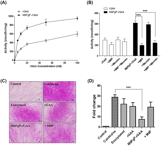
Enhanced uptake of M6PgP-conjugated rGAA by Pompe disease patient fibroblasts. (A) Intracellular GAA activities of Pompe disease patient fibroblasts were analyzed after incubation with increasing concentrations of rGAA (open circle) and M6PgP-conjugated rGAA (M6PgP-rGAA; closed circle). (B) The fibroblasts were incubated with 20 nM of rGAA (white bar) or M6PgP-rGAA (black bar) in the presence or absence of 5 mM M6P and/or 2 mg/ml mannan. Data are shown as mean ± standard deviation (n = 3). (C) Representative images of PAS-stained Pompe fibroblasts (magnification: x 400). Glycogens were accumulated in the cells cultured in the presence of colchicine (Colchicine). The accumulated glycogens slightly decreased through exocytosis in the absence of colchicine (Exocytosed) and were further digested by addition of 100 nM rGAA (rGAA) or M6PgP-conjugated rGAA (M6PgP-rGAA). Free M6Ps competitively inhibited glycogen clearance by M6PgP-conjugated rGAA (+M6P). Scale bar = 50 µm. (D) The images of PAS-stained cells in each well of 12-well culture plates were analyzed by using NIH Image J software (http://rsb.info.nih.gov/ij/). Strong purple spots over the arbitrary threshold value were identified and their areas were used for calculation of relative fold changes in comparison with the strong purple spot area of control cells (Supplementary Fig. S6). Data are shown as mean ± standard deviation (n = 4). One-way ANOVA with Tukey’s multiple comparison test, ***P < 0.001.

