Figure 3.

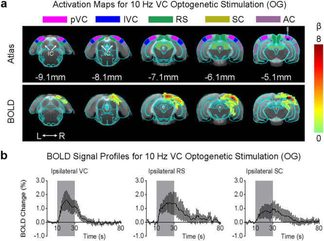
Optogenetic stimulation of the primary visual cortex at 10 Hz does not induce fMRI response in the IC. (a) Atlas (from the Paxinos & Watson) and activation (β) maps for the 10 Hz VC optogenetic stimulation (OG) overlaid on anatomical MRI. Activated voxels detected in the local VC, including pVC and lVC, RS, and ipsilateral SC and AC (n = 9; p < 0.05, corrected for FWE) are shown by the heat map. Abbreviations of atlas overlay are as follows: pVC (primary visual cortex), lVC (lateral visual cortex), RS (retrosplenial cortex), SC (superior colliculus), and AC (auditory cortex). (b) BOLD signal profiles in the significant voxels identified in (a). The results are presented as means ± standard error of the mean. Area in shade indicates the 20 s 10 Hz optogenetic stimulation.
