FIGURE 3.

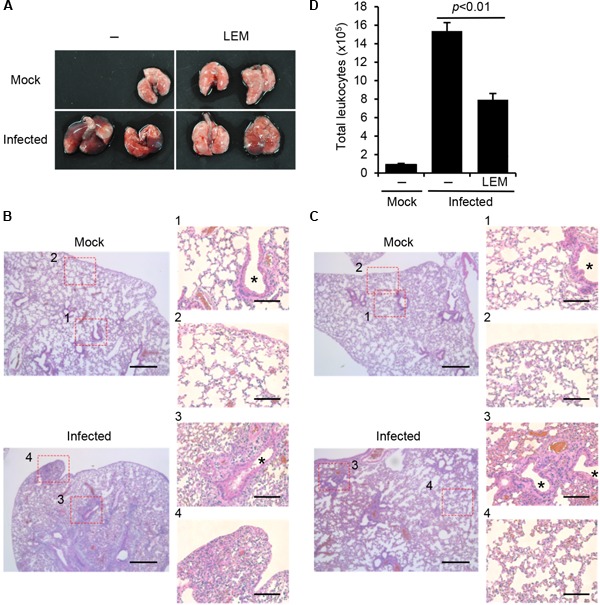
Effect of LEM on acute pulmonary influenza virus infection. Mice were intranasally infected with 1,000 pfu of PR8, and then orally administered with 1 mg/ml LEM. At 7 days post infection, the lungs of infected mice were dissected for pathological analysis. (A) shows the gross changes of the lungs. (B) (PBS) and (C) (LEM administration) show the hematoxylin and eosin-stained sections with enlarged figures. Asterisks indicate bronchioles. Lung section was obtained from two mice for every group and representative images are shown. Scale bar; 400 and 100 μm (in enlarged panels). In (D) bronchoalveolar lavage fluids (BALF) were obtained from the dissected lungs (n = 3 for mock-infected group; n = 3 for non-treated infected group; n = 3 for LEM-administrated infected group), and the number of leukocytes was counted by flow cytometry with anti-CD45.2 antibody, which is a leukocyte marker. The results are averages from three independent experiments with standard deviations, and the statistical significance was determined by Student’s t-test.
