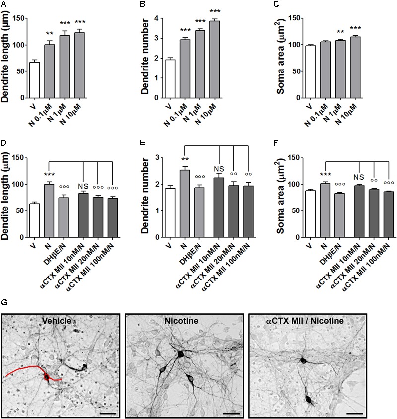
FIGURE 1.

Structural plasticity induced by nicotine in mouse DA neurons and blockade with α-conotoxin MII. (A–C) Concentration–response curves of the effect of nicotine (0.1–10 μM) on structural plasticity measured as maximal dendrite length, number of primary dendrites, and soma area. (D–F) Antagonism of the 10 μM nicotine-induced structural plasticity produced by pretreatment with 10–100 nM α-conotoxin MII; 10 μM dihydro-β-erythroidine was used as internal standard; (D) maximal dendrite length, (E) number of primary dendrites, and (F) soma area. (G) Representative photomicrographs of mouse mesencephalic DA neurons 72 h after exposure to vehicle, 10 μM nicotine, or 100 nM α-conotoxin MII followed by 10 μM nicotine. Red line drawing shows how the measurements of the three parameters, dendrite length, dendrite number, and soma area were performed (Scale bar: 50 μm). One-way ANOVA was used to analyze data in panels (A–C) and (D–F), respectively. Student’s t-test was used to compare dihydro-β-erythroidine with nicotine in (D–F). Data are expressed as mean ± SEM (∗∗∗p < 0.001; ∗∗p < 0.01 vs. vehicle; ∘∘∘p < 0.001; ∘∘p < 0.01 vs. nicotine; NS, non-significant, post hoc Bonferroni’s test). V: vehicle; N: nicotine; αCTX MII: α-conotoxin MII; DHβE: dihydro-β-erythroidine.
