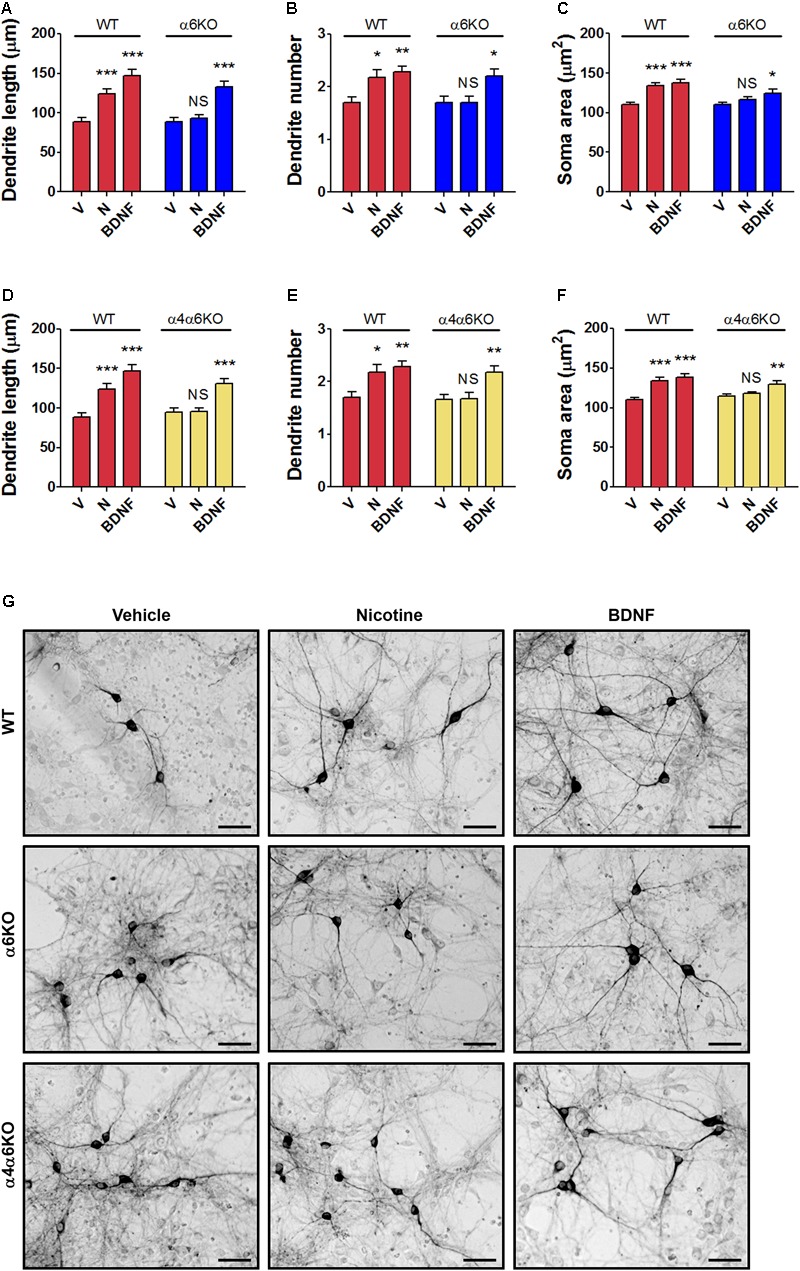
FIGURE 3.

Structural plasticity induced by nicotine in mesencephalic DA neurons from wild-type (WT) mice and α6 or α4/α6 nAChR subunit null mutant mice. Morphological effects of nicotine on DA neurons from (A–C) α6KO and WT mice and (D–F) α4/α6KO and WT mice on maximal dendrite length (A,D), number of primary dendrites (B,E) and soma area (C,F) measured 72 h after exposure to nicotine (10 μM), or BDNF (10 ng/ml) here used as active control group. Two-way ANOVA was used to analyze data in panels (A–C) and (D–F), respectively. Data are expressed as mean ± SEM (∗∗∗p < 0.001; ∗∗p < 0.01; ∗p < 0.05 vs. vehicle; NS, non-significant, post hoc Bonferroni’s test). (G) Representative photomicrographs of mouse mesencephalic DA neurons from WT, α6KO and α4/α6KO mice 72 h after exposure to vehicle, 10 μM nicotine or 10 ng/ml BDNF (Scale bar: 50 μm). WT: wild-type, V: vehicle, N: nicotine, BDNF: brain-derived neurotrophic factor.
