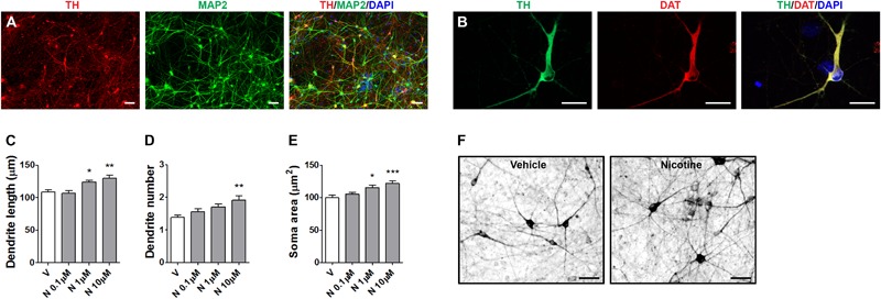
FIGURE 5.

Structural plasticity induced by nicotine in human iPSC-derived DA neurons. Representative images of dual immunofluorescence indicating coexpression of (A) TH (red) and MAP2 (green) and (B) TH (green) and DAT (red) assessed at day 70. Cell nuclei were stained with DAPI (blue). Scale bar: (B) = 50 μm; (C) = 30 μm. (C–E) Concentration–response of nicotine effects on structural plasticity measured as (C) maximal dendrite length, (D) number of primary dendrites, (E) soma area. One-way ANOVA was used to analyze data in panels (C–E). (F) Representative photomicrographs of human iPSC-derived DA neurons 72 h after exposure to vehicle or 10 μM nicotine (Scale bar: 50 μm). Data are expressed as mean ± SEM. (∗∗∗p < 0.001; ∗∗p < 0.01; ∗p < 0.05 vs. vehicle, post hoc Bonferroni’s test). V: vehicle, N: nicotine.
