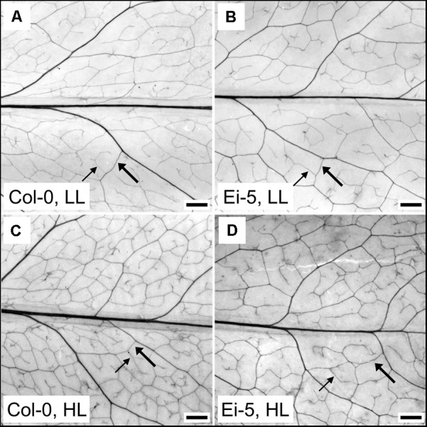
FIGURE 4.

Images of representative chemically cleared leaves of the (A,C) Col-0 and (B,D) low-vein-density Ei-5 accessions of A. thaliana grown under a 9-h photoperiod of (A,B) 100 (LL) or (C,D) 1000 (HL) μmol photons m-2 s-1 at a leaf temperature of 20°C. To better visualize the leaf vascular network, the contrast of each image was enhanced using the “Auto Contrast” feature in Adobe Photoshop CS4 (Adobe Photosystems, Inc., San Jose, CA, United States). Representative third (3°) and fourth (4°) order minor veins are indicated with thick and thin arrows, respectively. Scale bar = 1 mm.
