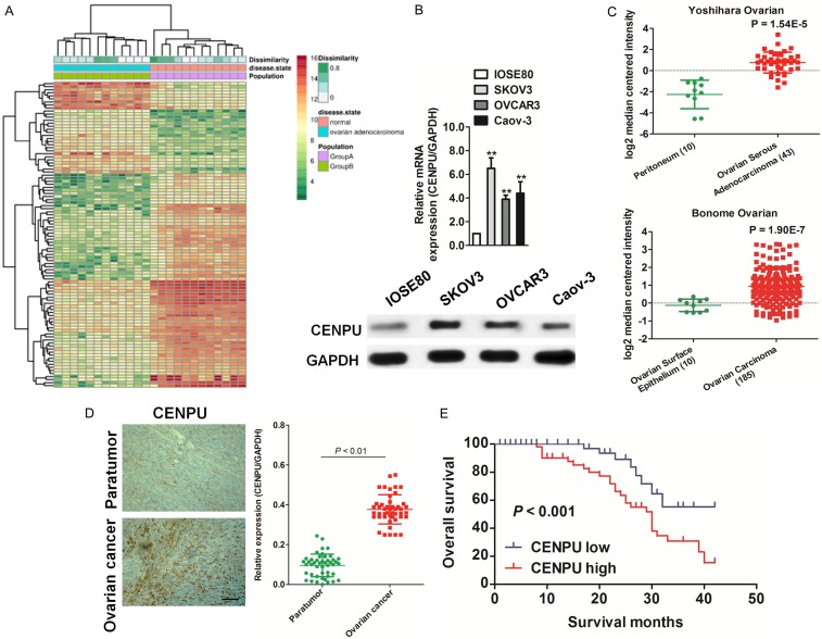
Figure 1.
The expression of CENPU in ovarian cancer. A. The expression profiling of mRNAs shown that CENPU was over-expressed in ovarian cancer as compared to normal (GDS3592). B. qRT-PCR and western blotting analyzed the expression of CENPU in IOSE80 cells and ovarian cancer cells. C. Box plots shown elevated levels of CENPU in ovarian cancer compared to normal from two microarray data-sets. **P < 0.01, compared with normal tissues. D. Representative staining result of CENPU in ovarian cancer tissues (left panel). Scale bar: 200 μm. qRT-PCR assay was conducted to analyze the expression of CENPU in ovarian cancer tissues. E. Kaplan-Meier survival curves of ovarian cancer patients with different expression level of CENPU.

