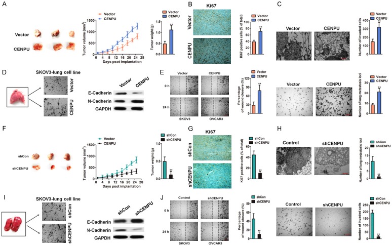
Figure 3.
Effect of CENPU on ovarian cancer growth and metastasis in vivo. A. Tumors were obtained four weeks after subcutaneous injection of CENPU over-expressing SKOV3 cells. Comparison of tumor volumes and weights between the CENPU over-expressing SKOV3 cells and control groups. B. The positive of Ki67 of tumor was determined in the experimental and control groups. Scale bar: 200 μm. C. Representative images of H&E-stained lungs with metastases. Scale bar: 200 μm. **P < 0.01 compared to vector. D. E-cadherin and N-cadherin were assessed by western blotting. E. Wound-closure analysis was performed to determine the effects of CENPU over-expressing SKOV3-lung cell line by calculating percentage of wound closure (left panel). CENPU over-expressing SKOV3-lung cell line was seeded onto Matrigel-coated Transwell chambers. After 24 h, the invaded cells were counted (right panel). Scale bar: 200 μm. **P < 0.01 compared to vector. F. Tumors were obtained after subcutaneous inoculation of shCENPU SKOV3 cells. Comparison of tumor volumes and weights between the shCENPU SKOV3 cells and control groups. G. The positive of Ki67 was evaluated in the experimental and control groups. H. Representative images of H&E-stained lungs with metastases. Scale bar: 200 μm. **P < 0.01 compared to shCon. I. E-cadherin and N-cadherin were determined by western blotting assay. J. Wound closure assay was performed to assess the effects of CENPU down-expressing SKOV3-lung cell line by calculating percentage of wound closure (left panel). CENPU down-expressing SKOV3-lung cell line was seeded onto Matrigel coated Transwell chambers. After 24 h, the invaded cells were counted (right panel). Scale bar: 200 μm. **P < 0.01 compared to shCon.

