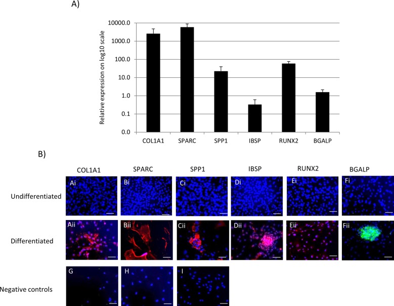
Fig. 1.
Osteoblast-associated gene and protein expression in equine iPSCs differentiated in osteogenic media on an OsteoAssay surface for 21 days. (A) Quantitative PCR analysis of osteoblast-associated genes. The mean of three independent replicates is shown. Error bars represent the s.e.m. and the relative expression (to the 18S housekeeping gene) is plotted on a log10 scale. (B) Immunocytochemistry is shown to detect osteoblast-associated proteins in undifferentiated iPSCs and iPSCs after 21 days of differentiation in osteogenic media. All proteins are detected following differentiation [(Ai–Eii) red staining; (Fi,Fii) green staining]. Negative controls for the secondary antibodies were performed on differentiated iPSCs (G) donkey anti-goat alexafluor 594. (H) Goat anti-rabbit alexafluor 594. (I) Goat anti-mouse FITC. DAPI staining of cell nuclei is shown in blue. Scale bars: 40 μm. Immunocytochemistry was performed on all lines of iPSCs and representative images are shown.

