Fig. 2.
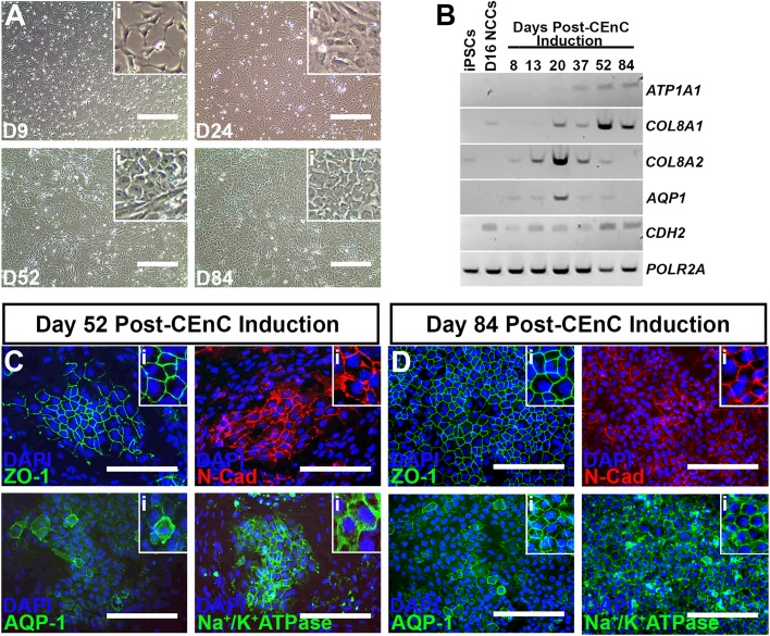
Differentiation of neural crest cells into corneal endothelial cells. (A) Representative light micrographs of neural crest cells differentiating into corneal endothelial cells at 9 (D9), 24 (D24), 52 (D52) and 84 (D84) days post-CEnC induction. Insets (i) in each panel show higher magnification views to better display changes in cell morphology throughout the differentiation process. (B) Rt-PCR for CEnC-specific transcripts comparing undifferentiated iPSCs and day 16 NCCs (D16 NCCs) to differentiated cells at 8, 13, 20, 37, 52 and 84 days post-CEnC induction. POLR2A served as a control transcript. (C) Representative immunocytochemical labeling of differentiated cells at 52 days post-CEnC induction with the CEnC-specific markers, zonula occludens-1 (upper left; ZO-1, green), N-Cadherin (upper right; N-Cad, red), Aquaporin-1 (lower left; AQP-1, green) and Na+/K+ATPase (lower right, green). DAPI was used to counterstain cell nuclei. Insets (i) in each panel show higher magnification views. (D) Representative immunocytochemical labeling of differentiated cells at 84 days post-CEnC induction with ZO-1 (upper left, green), N-Cad (upper right, red), AQP-1 (lower left, green) and Na+/K+ATPase (lower right, green). DAPI was used to counterstain cell nuclei. Insets (i) in each panel show higher magnification views. Scale bars: 400 µm in A; 200 µm in C,D.

