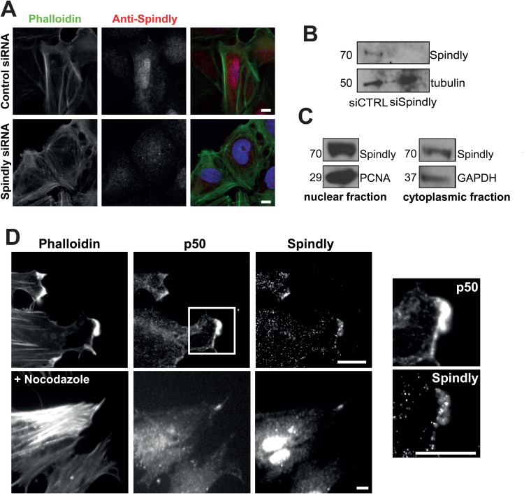
Fig. 1.
Spindly localizes to the leading edge of fixed migrating cells. (A) Confluent U2OS cells were treated with control or Spindly-specific siRNAs and then cells were fixed and stained to visualize nuclei (DAPI), filamentous actin (phalloidin) and Spindly. (B) An immunoblot of cell lysates show that Spindly was efficiently depleted by the siRNAs. (C) U2OS cells were lysed and the cytoplasmic and nuclear fractions were separated. Co-fractionation with PCNA confirms Spindly presence in the nucleus and co-fractionation with GAPDH confirms the presence of Spindly in the cytoplasm. (D) Foreskin fibroblasts were cultured to confluency, and then the monolayer was scratched to promote cell migration. 4 h after scratch-wounding, cells were fixed and stained to visualize filamentous actin (phalloidin), p50 Dynamitin, and Spindly. Images on the left show a magnification of the box shown in the upper image. Nocodazole treatment did not abolish the colocalization of p50 and Spindly. Scale bars: 10 µm.

