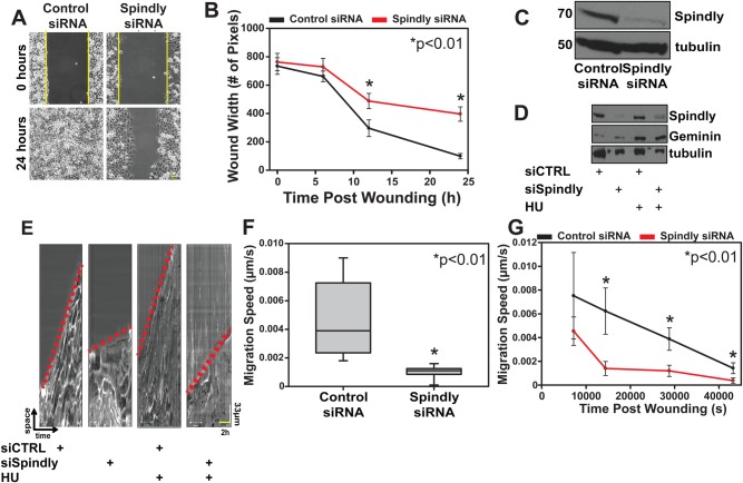
Fig. 3.
Spindly is required for rapid cell migration. (A,B) U2OS cells treated with control or Spindly-specific siRNAs were plated into an ibidi silicone culture-insert inside an imaging chamber. After cells reached confluency, the insert was removed and the closure of the induced wound was followed over time using phase-contrast microscopy. Three independent experiments were performed and data in the graph represent mean±s.d. (C) Western blotting confirmed that the Spindly-specific siRNAs were effective at depleting the protein. (D) Spindly siRNA and control siRNA treated cells were treated with hydroxyurea (HU) to block DNA synthesis and arrest cells at the entry of S-phase. Geminin protein levels confirmed that the HU treatment worked. Tubulin used as loading control. (E–G) Kymographs generated from control and Spindly-depleted cells migration movies. Red dotted lines to indicate the different slopes (i.e. velocity) between control and silenced cells. The kymographs were used to measure the speed of cells at various time points post wounding. The Spindly-silenced cells were slower to migrate into a wound, regardless of whether or not HU was added to the cells. Three independent experiments were performed and data in the graph represent mean±s.d. Student's t-test was used to determine the statistical significance.

