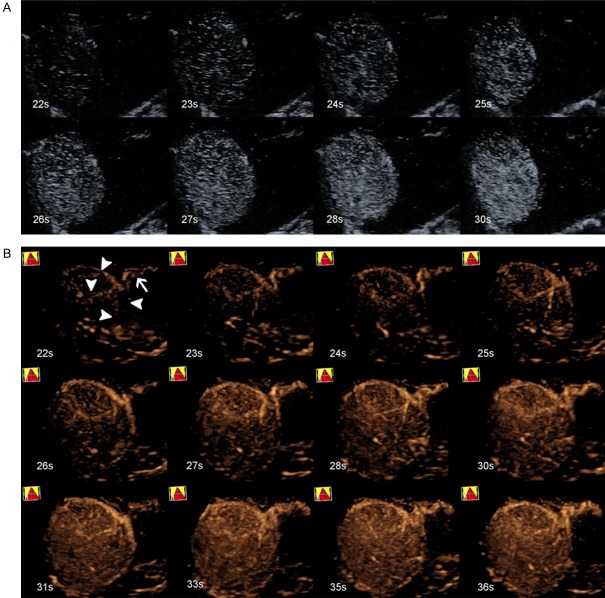
Figure 1.
A 68-year-old male with HCC (tumor size, 8.1 cm × 5.6 cm) in segments V and VIII of the liver. A. 2D-CEUS did not exhibit the feeding artery during the whole process. B. Real-time 3D-CEUS showed a clear feeding artery (white arrow), with four surrounding branches (white arrowhead) supplying the lesion, which is called a surrounding pattern.

