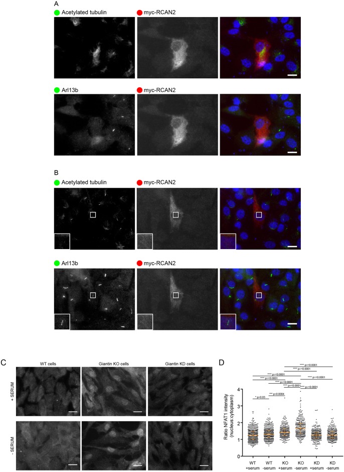
Fig. 4.
Giantin KO cells show higher levels of NFAT1 activation. (A) Expression of myc-RCAN2 (red in merge) prevents cilia formation as shown by acetylated tubulin or Arl13b (both pseudocoloured green in merge; images were obtained from cells triple labelled with Alexa Fluor 488, 568 and 647). 80% of myc-RCAN2-expressing cells failed to extend a cilium (n=100). (B) In 5% of these myc-RCAN2-expressing cells, we observed ‘decapitated’ cilia, positive for Arl13b but not for acetylated tubulin. (C) Representative images showing WT and giantin KO and knockdown (KD) cells grown in serum or serum deprived for 24 h and then immunolabelled for NFAT1. Scale bars: 10 μm. (D) Quantification of the ratio of nuclear to cytoplasmic NFAT1 staining intensity in experiments represented in A [n=3, bars represent median and interquartile range, P-values calculated using non-parametric ANOVA (Kruskal-Wallis test with Dunn's multiple comparisons test)].

