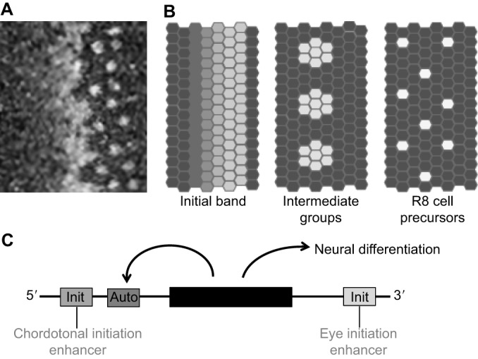
Fig. 2.

Regulation of Atonal expression and transcription in the Drosophila eye. (A) Confocal image of Atonal (Ato) protein expression at the leading edge of the Drosophila retina differentiation wave. Towards the anterior (left), Ato expression progressively accumulates in all the cells, then rapidly evolves through a pattern of intermediate groups of cells that transiently maintain Ato protein while the surrounding cells lose expression, resolving to only isolated single cells that maintain Ato expression posteriorly (right). These are now the committed and postmitotic R8 photoreceptor precursors. The wave of differentiation advances anteriorly at a rate of one column every 90-100 min. (B) The complete Ato expression pattern can be dissected into at least three temporally overlapping phases (Jarman et al., 1995). First, Ato expression accumulates uniformly, reaching higher and higher levels more posteriorly until this uniform expression abruptly ceases. Replacing the uniform expression, and just overlapping with it in time, is a transient phase of expression in ‘intermediate groups’ of up to ten cells. The intermediate groups are proneural preclusters that will all develop as R8 photoreceptor neurons unless Notch signaling is activated (Baker et al., 1996; Dokucu et al., 1996). Then, each intermediate group is refined to a single Ato-expressing cell that maintains expression for three to four ommatidial columns (∼6 h). (C) Uniform initiation of Ato expression depends on an eye initiation enhancer that is downstream of the coding region of the ato gene (black box), but is independent of functional Ato protein and of the 5′ autoregulatory enhancer. By contrast, expression in the intermediate groups of up to ∼10 cells is autoregulatory and depends on the 5′ autoregulatory enhancer because the 3′ initiation enhancer becomes inactive at this stage. Single cells escape Notch activity to maintain autoregulatory Ato expression and become determined as R8 photoreceptor precursor cells. They maintain Ato expression for ∼6 h, then Ato protein undergoes inhibitory phosphorylation, leading to the loss of expression. This loss is permanent because initiation is no longer active. Other neural regions that express Ato (e.g. chordotonal organs) rely on distinct enhancers (Baker et al., 1996; Sun et al., 1998; Quan et al., 2016).
