
Fig. 3.
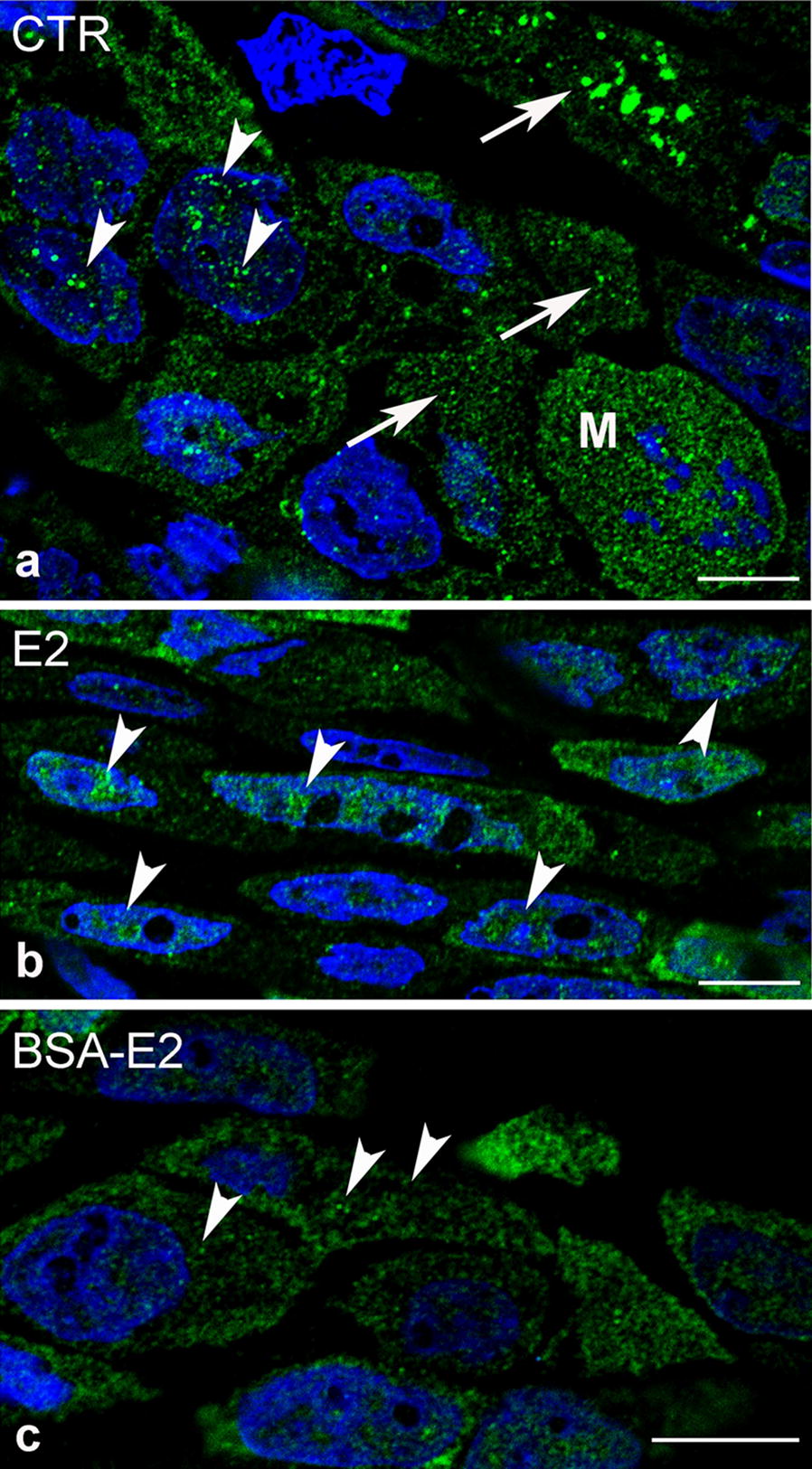
ER-α labeling of control and treated MCF-7 cells on semithin frozen sections. a ER-α receptor labeling occurred in aggregates or as punctate structures both inside the nucleus (arrowheads) as well as in the cytoplasm of untreated MCF-7 cells. Observe a mitotic form (M) where intensive ERα expression could be detected. b Upon E2 treatment (2 min, 10−8 M/L), the majority of receptor labeling accumulated inside the nucleus (arrowheads). c Immunofluorescence labeling of ER-α could predominantly be observed in the cytoplasm and submembranous localization of MCF-7 cells upon BSA-E2 treatment (arrowheads). Nuclei were stained with DAPI, bars indicate 10 µm