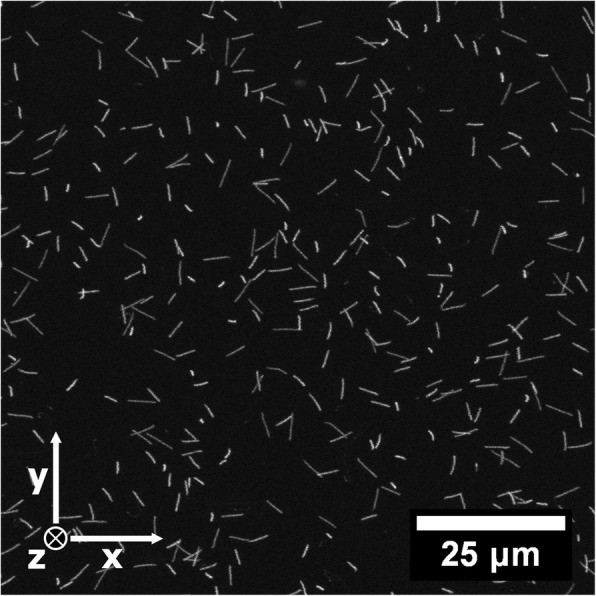
Fig. 1.

A 2D maximum intensity projection of the fluorescence measured in a 100 × 100 × 12 μm3 (x,y,z) volume with a confocal laser scanning microscope in a FNTD after irradiation with alpha particles. The apparent length of the tracks is related to the angle of incidence of the alpha particles. Individual tracks were analyzed using in-house build software, yielding the start- and endpoint, direction, relative scattering, fluorescence intensity and energy for each track [20]
